


Microfracture surgery is a commonly used arthroscopic technique for treating localized cartilage defects, especially in the knee joint. Cartilage injuries can cause pain, swelling, reduced mobility, and mechanical symptoms that may affect daily life and athletic performance.
Because articular cartilage has limited healing capacity, orthopedic surgeons use different techniques to stimulate biological repair. Microfracture is one of the most established methods used to encourage the formation of new repair tissue in damaged cartilage areas.
This guide explains how microfracture surgery works, when it is used, and why proper arthroscopic instruments are important for successful cartilage repair.
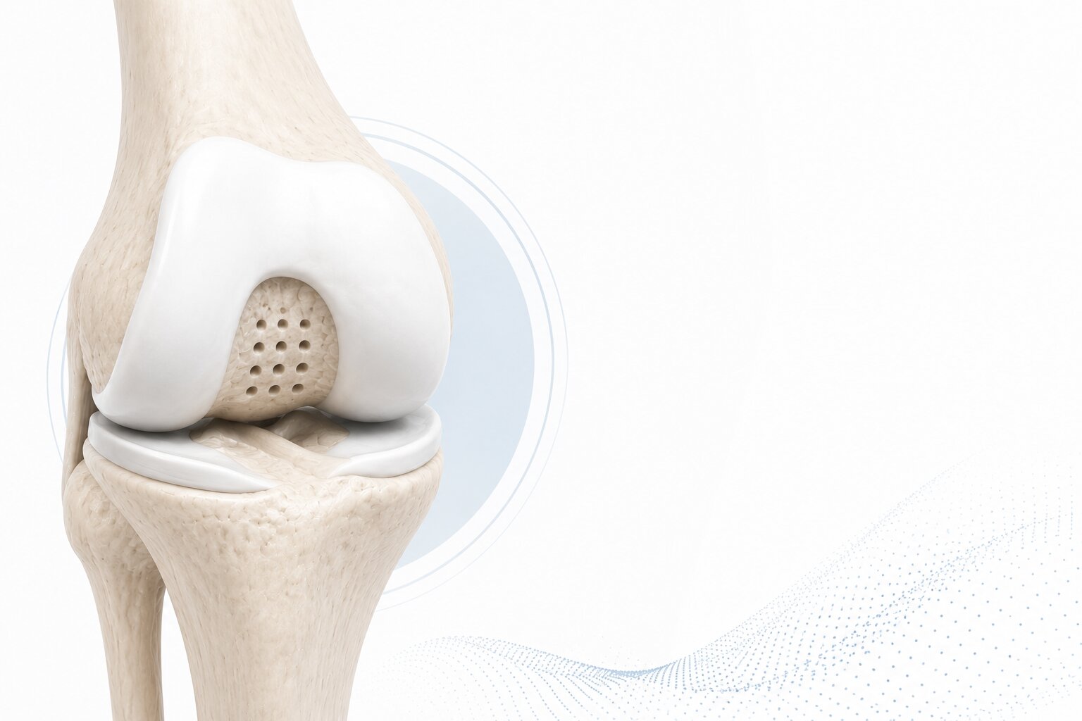
Articular cartilage is the smooth, protective tissue covering the ends of bones inside a joint.
Its main functions include:
Healthy cartilage allows the joint to move smoothly and painlessly. However, cartilage can become damaged due to trauma, sports injuries, degeneration, or repetitive overload.
Unlike many other tissues, articular cartilage has very limited blood supply.
This means it has poor natural healing potential.
When cartilage is damaged, the body cannot easily repair the defect on its own. Untreated cartilage defects may lead to:
For this reason, cartilage repair techniques are important in modern orthopedic surgery.
Microfracture surgery is an arthroscopic cartilage repair technique.
During the procedure, the surgeon creates small holes in the subchondral bone beneath the cartilage defect. These small perforations allow bone marrow elements to enter the damaged area.
The marrow contains cells and biological factors that support the formation of repair tissue.
The goal is to cover the cartilage defect with fibrocartilage-like repair tissue.
The principle of microfracture is based on biological stimulation.
After the damaged cartilage area is prepared, small channels are created in the underlying bone.
This allows:
Over time, this clot matures into fibrocartilage-like tissue that helps fill the defect.
Microfracture is commonly considered for selected patients with focal cartilage defects.
It may be used in cases involving:
Patient selection is very important.
Microfracture is generally more suitable when the defect is localized rather than diffuse degenerative cartilage loss.
Microfracture is most frequently performed in the knee, but it may also be used in other joints.
Common areas include:
The knee remains the most common joint for microfracture procedures.
The procedure begins with arthroscopic evaluation of the joint.
The surgeon assesses:
This evaluation helps determine whether microfracture is appropriate.
The damaged cartilage area is carefully prepared.
Loose or unstable cartilage fragments are removed.
The goal is to create stable cartilage borders around the defect.
Proper preparation is essential because unstable edges may compromise repair tissue formation.
In many cases, the calcified cartilage layer must be removed to expose the subchondral bone.
This step supports better access to marrow elements.
Using a microfracture awl, pick, or specialized microfracture system, the surgeon creates small holes in the subchondral bone.
The holes should be:
Precision is critical.
After creating the holes, the surgeon confirms that marrow elements are entering the defect area.
This indicates that biological stimulation has been achieved.
After surgery, a blood clot forms within the cartilage defect.
This clot becomes the foundation for fibrocartilage repair tissue.
Microfracture offers several advantages in selected patients.
The procedure is performed arthroscopically through small portals.
This reduces soft tissue trauma compared to open surgery.
Microfracture uses the body’s own marrow elements to stimulate repair.
Compared with more complex cartilage restoration procedures, microfracture is relatively simple and cost-effective.
When properly indicated, microfracture can help reduce symptoms and improve function.
Microfracture is not ideal for every cartilage problem.
Potential limitations include:
Correct patient selection remains essential.
Other cartilage repair options include:
Microfracture remains widely used because it is technically accessible and can be performed during arthroscopy.
However, more advanced techniques may be preferred for larger or complex defects.
Microfracture success depends heavily on precise instrumentation.
The instrument must allow controlled penetration into subchondral bone while minimizing surrounding cartilage damage.
Modern microfracture systems are designed to provide:
Specialized microfracture instruments help surgeons perform the technique more efficiently and consistently.
Rehabilitation is one of the most important parts of recovery.
Because repair tissue forms gradually, the joint must be protected during early healing.
Protocols may include:
Recovery time varies depending on defect size, location, and patient activity level.
Several factors influence microfracture outcomes:
Younger patients with small, contained lesions often show better results.
Orthosyn Medikal provides arthroscopic solutions designed to support modern cartilage repair procedures.
Microfracture systems are used to assist surgeons in creating controlled perforations in subchondral bone during arthroscopic cartilage repair.
Together with other arthroscopy products, microfracture instruments support minimally invasive orthopedic procedures and sports medicine applications.
Microfracture surgery is an established arthroscopic cartilage repair technique used for selected focal cartilage defects. By creating controlled perforations in subchondral bone, the procedure stimulates marrow-based repair and supports fibrocartilage formation.
Although microfracture is not suitable for every cartilage lesion, it remains an important option in modern orthopedic and sports medicine surgery.
Successful outcomes depend on proper patient selection, accurate technique, reliable instrumentation, and structured rehabilitation.