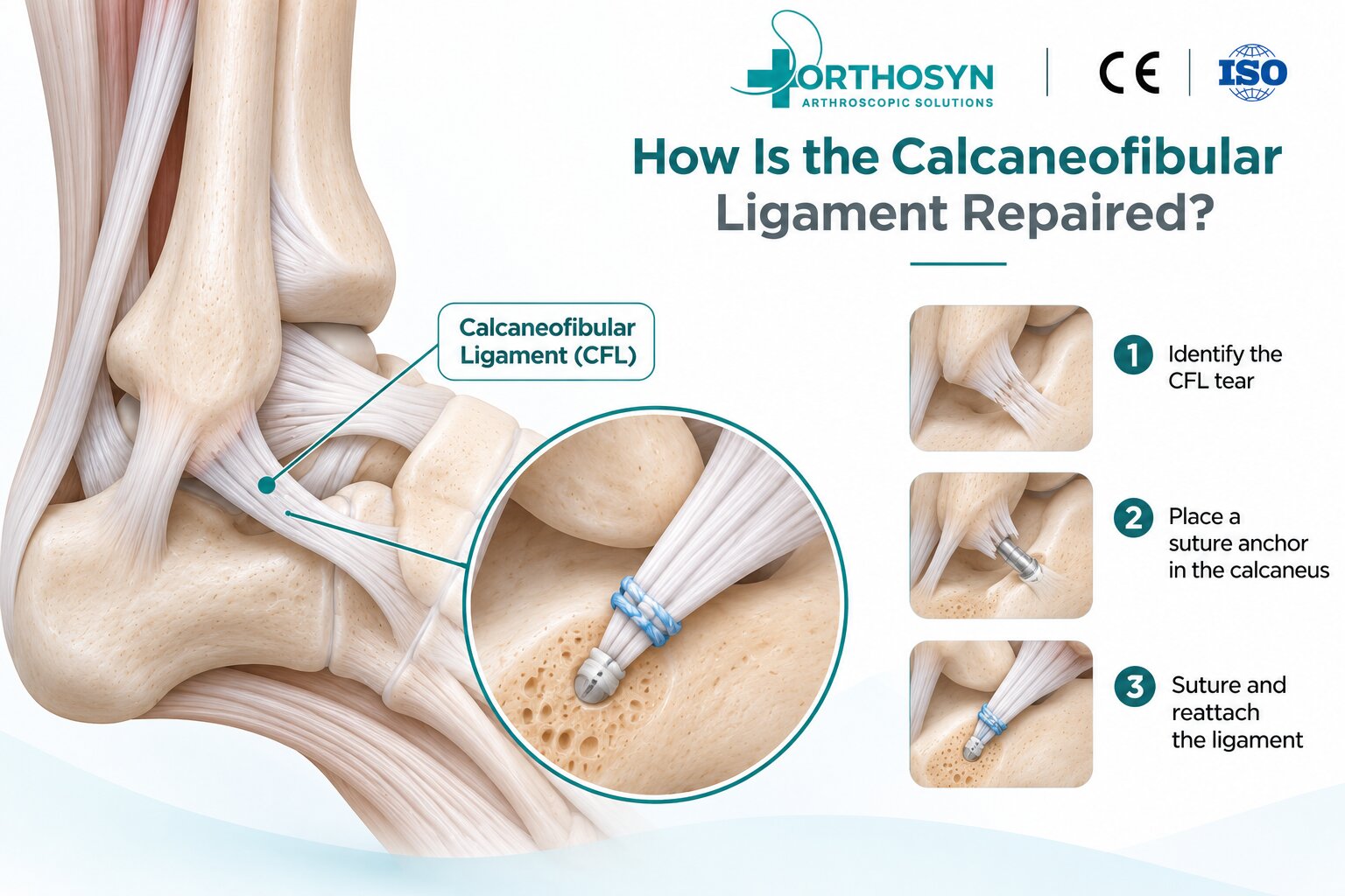
Calcaneofibular ligament repair is a surgical procedure used to restore lateral ankle stability when the calcaneofibular ligament (CFL) is damaged or insufficient. The CFL is one of the primary stabilizing ligaments on the outer side of the ankle and plays an important role in controlling inversion and subtalar joint movement.
CFL injuries frequently occur alongside anterior talofibular ligament (ATFL) injuries following ankle sprains. While many lateral ankle ligament injuries can be treated conservatively, surgery may be considered for persistent mechanical instability or selected severe injuries.
The fundamental repair concept is:
CFL Injury → Ligament Assessment → Tissue Preparation → Suture Fixation → Anatomical Reattachment → Stability Assessment
The calcaneofibular ligament connects the distal fibula to the lateral surface of the calcaneus.
It extends downward and backward from the lateral malleolus, crossing both the ankle and subtalar joints.
Its primary functions include:
Resisting excessive ankle inversion
Supporting lateral ankle stability
Contributing to subtalar joint stability
Restricting abnormal hindfoot movement
The CFL works together with the ATFL and posterior talofibular ligament (PTFL) to stabilize the lateral ankle.
The CFL is commonly injured during a forceful inversion ankle sprain.
Contributing factors include:
Sports-related ankle sprains
Sudden changes of direction
Landing awkwardly after jumping
Recurrent ankle instability
Falls or traumatic ankle inversion
Combined ATFL and CFL injuries
Isolated CFL injuries are less common than combined lateral ligament injuries.
Patients may experience lateral ankle pain, swelling, tenderness below the lateral malleolus, and a sensation that the ankle is giving way.
Chronic CFL insufficiency may contribute to recurrent ankle sprains and instability during walking on uneven surfaces.
Clinical examination may include the talar tilt test, assessment of the ATFL, and evaluation of subtalar stability. MRI or ultrasound can help assess ligament integrity when indicated.
CFL repair may be performed through an open, mini-open, or selected arthroscopic-assisted approach.
The surgeon evaluates the CFL injury, remaining ligament tissue, and associated lateral ankle structures.
The ATFL, peroneal tendons, and subtalar joint may also be examined.
The damaged ligament attachment is identified.
For an avulsion from the fibular attachment, the distal fibula may be prepared to provide a suitable surface for ligament reattachment.
The repair strategy depends on the injury location and available tissue quality.
When anchor fixation is indicated, a suture anchor may be inserted at the appropriate anatomical attachment site.
For many proximal CFL avulsion repairs, this involves the fibular attachment near the lateral malleolus.
The surgeon must account for the limited dimensions of the distal fibula and surrounding anatomical structures.
High-strength sutures are passed through the remaining CFL tissue.
The sutures are used to approximate the ligament to its anatomical attachment.
If the tissue is insufficient for direct repair, ligament reconstruction or augmentation may be considered instead.
The sutures are secured to restore ligament continuity and appropriate tension.
The repair should provide stability without excessively restricting physiological ankle or subtalar movement.
The surgeon evaluates ankle inversion stability, ligament tension, and joint movement.
Associated ATFL insufficiency may also require repair.
Characteristic | CFL Repair | ATFL Repair |
|---|---|---|
Ligament | Calcaneofibular ligament | Anterior talofibular ligament |
Anatomical connection | Fibula to calcaneus | Fibula to talus |
Main function | Inversion and subtalar stability | Anterior talar translation and rotational stability |
Common injury | Combined lateral ligament injury | Commonly injured during ankle sprains |
Fixation | Sutures or anchors when indicated | Sutures or anchors when indicated |
Both ligaments may be repaired during lateral ankle stabilization procedures, including modified Broström-type repairs.
Suture anchors may be used to secure the damaged ligament to bone.
The fixation construct typically involves:
Fibular Bone → Suture Anchor → High-Strength Suture → CFL Tissue
The suitability of an anchor depends on:
Available bone stock
Ligament tissue quality
Injury location
Anchor diameter
Insertion angle
Required fixation strength
Not every CFL injury requires an anchor, and not every anchor configuration is suitable for the distal fibula.
ORTHOSYN provides different technologies for orthopedic soft tissue-to-bone fixation.
Available diameters include 1.3 mm, 1.5 mm, 1.9 mm, and 3.0 mm.
These UHMWPE-based all-suture anchors offer small-diameter fixation options for compatible orthopedic procedures.
ORTHOSYN provides titanium anchors in several diameters, including 2.0 mm and 3.5 mm configurations.
PEEK anchor systems provide nonmetallic rigid fixation options for selected soft tissue repair procedures.
The use of any ORTHOSYN anchor for CFL repair must be supported by its product-specific indications, anatomical compatibility, and instructions for use.
Direct ligament repair and ligament reconstruction are different surgical strategies.
CFL Repair uses the remaining native ligament tissue to restore its anatomical attachment.
CFL Reconstruction uses a tendon graft or other reconstructive technique when the native ligament is insufficient for reliable repair.
Reconstruction may be considered in cases involving severe tissue degeneration, previous failed repair, or complex chronic instability.
The decision depends on tissue quality, ankle anatomy, and the patient's functional requirements.
Important factors include ligament tissue quality, anatomical reattachment, fixation stability, associated ATFL injury, hindfoot alignment, and rehabilitation.
Generalized ligamentous laxity or significant hindfoot varus may influence treatment planning and long-term stability.
Successful repair requires both mechanical support and biological healing.
Recovery depends on the repair technique, associated injuries, and rehabilitation protocol.
Initial rehabilitation generally includes a period of immobilization or protected weight-bearing, followed by progressive mobility, strengthening, and balance training.
Return to demanding sports may require approximately three to six months or longer, depending on functional recovery and surgical recommendations.
Calcaneofibular ligament repair aims to restore the anatomical continuity and stabilizing function of the CFL after lateral ankle injury.
The procedure may involve direct suturing, suture anchor fixation, or combined lateral ligament repair.
When native ligament tissue is insufficient, reconstruction may be necessary.
ORTHOSYN offers soft, titanium, and PEEK anchor technologies for compatible orthopedic soft tissue fixation applications.
Successful CFL repair depends on accurate anatomical positioning, appropriate fixation, tissue quality, and structured rehabilitation.
Explore the ORTHOSYN arthroscopy portfolio.