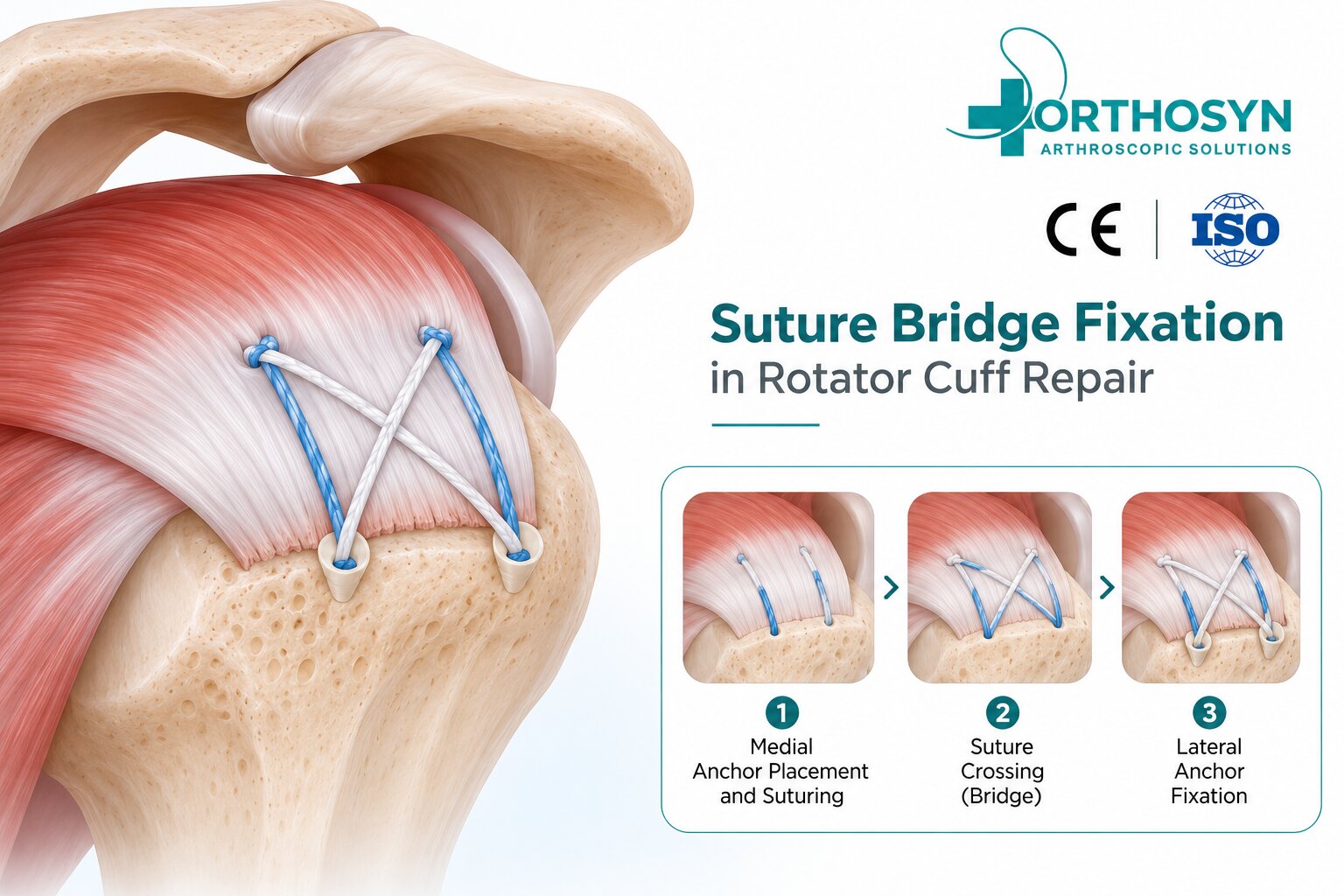
Suture bridge fixation in rotator cuff repair is an arthroscopic technique used to reattach torn rotator cuff tendons to the greater tuberosity of the humerus. It is commonly associated with a transosseous-equivalent double-row repair configuration, in which sutures extend from medial-row anchors to lateral-row fixation points.
The technique aims to provide broad tendon-to-bone contact, distribute compression across the repaired tendon, and maintain mechanical stability during biological healing.
The fundamental fixation concept is:
Rotator Cuff Tear → Footprint Preparation → Medial Anchor Placement → Suture Passage → Lateral-Row Fixation → Tendon Compression
Suture bridge fixation is a repair configuration that uses sutures spanning across the surface of a torn rotator cuff tendon.
In a typical construct, medial-row anchors are inserted near the articular margin of the greater tuberosity. Sutures are passed through the tendon and secured laterally using additional anchors, frequently knotless anchors.
This creates a bridging configuration that compresses the tendon against its anatomical insertion area, known as the rotator cuff footprint.
Suture bridge repair is frequently used for supraspinatus tendon tears and may be adapted for selected larger or multi-tendon rotator cuff injuries.
The technique uses two fixation regions:
Medial row: Anchors positioned closer to the articular margin provide initial suture fixation through the tendon.
Lateral row: Anchors positioned farther laterally secure the bridging sutures and create compression across the repair footprint.
The mechanical principle is:
Medial Suture Anchors → Tendon Sutures → Bridging Compression → Lateral Fixation Anchors
The objective is to maintain tendon approximation over a broad contact area while healing occurs at the tendon-bone interface.
The surgeon examines the rotator cuff tear, tendon mobility, tissue quality, and surrounding shoulder structures.
The tear pattern and degree of tendon retraction help determine whether a suture bridge configuration is appropriate.
The greater tuberosity footprint is prepared to create a suitable surface for tendon reattachment.
The surgeon removes interfering tissue as needed while preserving appropriate bone stock.
Suture anchors are positioned along the medial region of the prepared footprint.
The number and location of anchors depend on the tear dimensions, tendon characteristics, and selected technique.
High-strength sutures or suture tapes are passed through the torn rotator cuff tendon.
Suture positioning is planned to distribute loads and reduce excessive stress concentrations.
The sutures are brought across the tendon and secured using lateral-row anchors.
Knotless anchors are frequently used for this stage because they can secure the bridging sutures without requiring additional external knots.
The surgeon evaluates tendon approximation, footprint coverage, suture tension, and fixation stability.
The construct should provide appropriate compression without excessive tension that could compromise the tendon.
Technique | Fixation principle |
|---|---|
Single-Row Repair | Anchors secure the tendon along one row |
Conventional Double-Row Repair | Separate medial and lateral fixation rows support tendon reattachment |
Suture Bridge Repair | Sutures bridge between medial and lateral anchors, compressing the tendon over the footprint |
Suture bridge fixation is generally considered a form of double-row, transosseous-equivalent repair.
Biomechanical studies have demonstrated advantages in footprint contact characteristics and construct properties for some suture bridge configurations. However, improved biomechanical measurements do not automatically translate into superior clinical outcomes for every patient.
Knotless anchors are commonly selected for lateral-row fixation.
Potential technical advantages include:
Securing bridging sutures without additional knot tying
Intraoperative adjustment of suture tension
Controlled compression across the tendon footprint
Compatibility with selected suture tape configurations
Flexible positioning of lateral fixation points
The performance of a knotless construct depends on the anchor design, bone quality, suture material, and implantation technique.
Knotless fixation is not universally superior to knotted fixation.
ORTHOSYN provides several anchor technologies relevant to arthroscopic soft tissue-to-bone fixation.
Available in 3.5 mm and 5.0 mm configurations, these anchors use PEEK components within a knotless fixation system.
These systems combine PEEK and titanium components and are available in 3.5 mm and 5.0 mm configurations.
ORTHOSYN also offers conventional suture anchors that may be considered for compatible medial-row fixation applications.
High-strength UHMWPE-based sutures support different orthopedic soft tissue fixation constructs.
The availability of these components does not establish that every combination forms a validated suture bridge system. Anchor compatibility, permitted suture configurations, anatomical indications, and the instructions for use must be verified before selecting a specific construct.
Important factors include:
Rotator cuff tear size and pattern
Tendon thickness and tissue quality
Bone density of the greater tuberosity
Anchor placement and pullout resistance
Suture and suture tape characteristics
Medial-row tissue loading
Lateral-row fixation stability
Appropriate tension across the repair
Excessive compression or medial-row tension may contribute to tissue damage in susceptible repairs.
Medial-row failure describes a retear occurring near the medial fixation region while portions of the lateral tendon attachment may remain intact.
This failure pattern has been described following some double-row and suture bridge repairs.
Potential contributing factors include high medial-row tension, compromised tendon tissue, tear characteristics, and biological healing limitations.
The technique should therefore aim to balance broad footprint compression with appropriate tissue tension.
Potential advantages of suture bridge fixation include broader footprint compression, multiple fixation points, and favorable initial biomechanical properties in selected constructs.
However, the technique may require additional anchors, careful suture management, and greater attention to tendon tension.
Not every rotator cuff tear is suitable for a double-row or suture bridge repair. Large retracted tears, poor tissue quality, or limited bone stock may require alternative strategies.
Rehabilitation depends on the size of the tear, tendon quality, repair stability, and associated procedures.
Initial treatment often includes sling protection and restricted active shoulder loading. Controlled movement and progressive strengthening are introduced according to the surgeon's protocol.
Return to demanding physical activity may require six months or longer, while tendon-to-bone healing and functional recovery vary between patients.
Suture bridge fixation in rotator cuff repair is an arthroscopic double-row technique designed to maintain tendon-to-bone contact using sutures spanning between medial and lateral fixation anchors.
The technique combines medial-row suture fixation with lateral-row compression to support tendon reattachment at the greater tuberosity footprint.
ORTHOSYN offers PEEK, titanium, and knotless anchor technologies, together with high-strength sutures, for compatible orthopedic soft tissue fixation procedures.
Successful repair depends on appropriate implant selection, anatomical placement, balanced suture tension, biological healing, and structured rehabilitation.
Explore the ORTHOSYN arthroscopy portfolio.