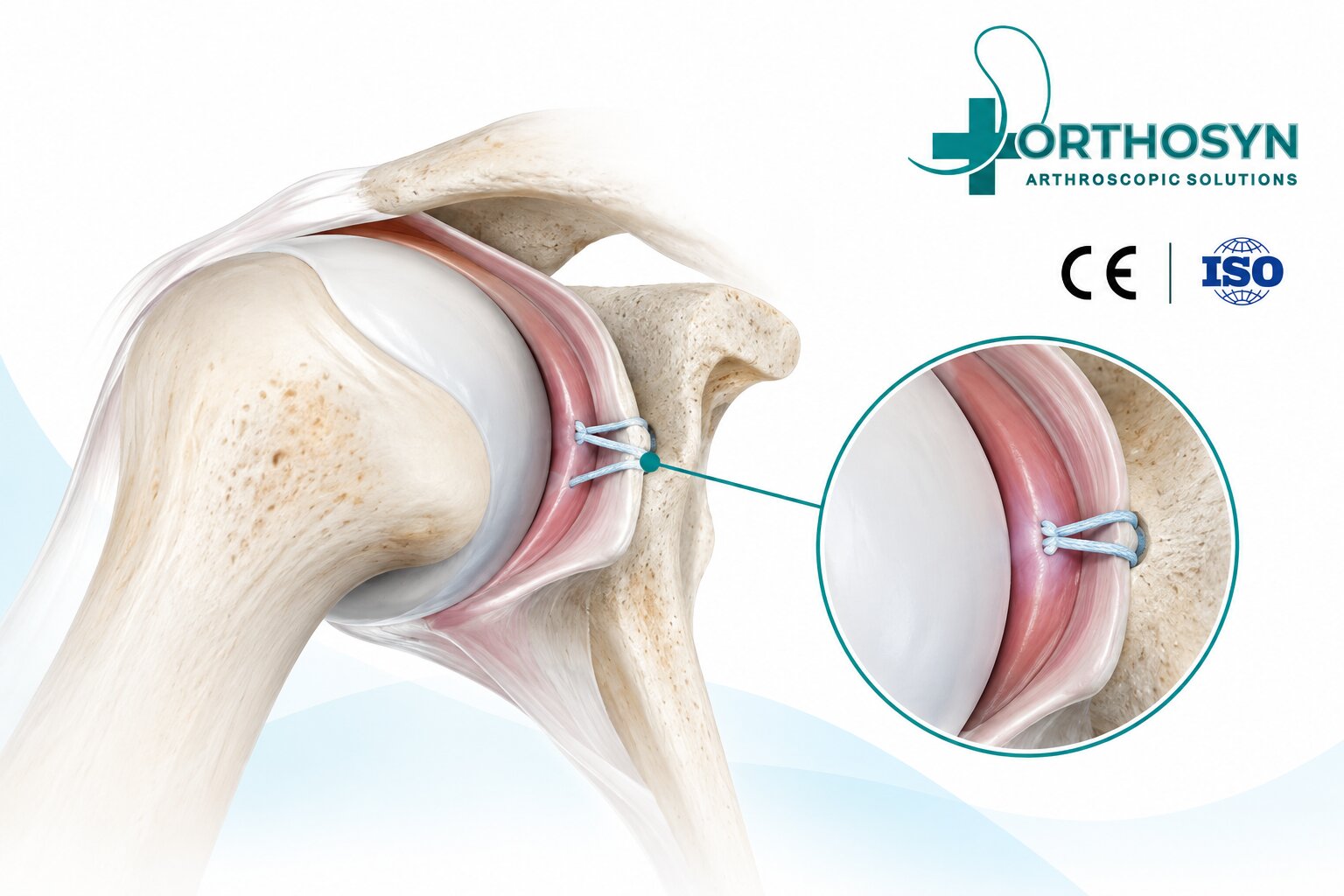
Suture anchor fixation in posterior labral repair is an arthroscopic technique used to restore the attachment of the posterior glenoid labrum and improve shoulder stability. Posterior labral injuries may occur following trauma, repetitive loading, or sports activities that place excessive stress on the shoulder joint.
The posterior labrum helps stabilize the humeral head within the glenoid socket. When damaged, patients may experience pain, clicking, weakness, or recurrent posterior shoulder instability.
Arthroscopic posterior labral repair uses specialized sutures and bone anchors to reattach the damaged labrum to the glenoid rim.
The fundamental repair concept is:
Posterior Labral Tear → Glenoid Preparation → Suture Anchor Placement → Labral Reattachment → Shoulder Stabilization
The glenoid labrum is a fibrocartilaginous structure surrounding the glenoid cavity of the scapula.
It increases the effective depth of the shoulder socket and contributes to joint stability.
The posterior labrum is located along the back portion of the glenoid rim and helps resist posterior translation of the humeral head.
Posterior labral injuries can compromise this stabilizing function, particularly during activities involving pushing, lifting, or repetitive shoulder loading.
Posterior labral tears can result from acute trauma or repetitive mechanical stress.
Common causes include:
Falls onto an outstretched arm
Posterior shoulder dislocation or subluxation
Contact sports
Weightlifting and bench pressing
Repetitive overhead activities
Traumatic loading of the shoulder
Chronic posterior shoulder instability
Some posterior labral tears occur together with posterior capsular injuries or glenoid bone abnormalities.
Symptoms vary depending on the injury and degree of instability.
Patients may experience:
Deep posterior shoulder pain
Clicking or catching sensations
Pain during pushing movements
Reduced shoulder strength
Difficulty with weightlifting
Recurrent shoulder subluxation
A sensation of shoulder instability
Clinical examination and imaging, including MRI or MR arthrography when appropriate, help identify the injury.
Arthroscopic posterior labral repair generally involves several stages.
The surgeon examines the glenohumeral joint to identify the location and extent of the posterior labral tear.
Associated cartilage damage, capsular laxity, and bone abnormalities are also evaluated.
The detached labral tissue is carefully mobilized.
The posterior glenoid rim is prepared to create a suitable surface for tissue reattachment and biological healing.
One or more suture anchors are inserted along the posterior glenoid rim.
Anchor positioning depends on the tear location, available bone, tissue quality, and selected repair technique.
Smaller-diameter anchors may be useful where bone preservation and limited anatomical space are important considerations.
High-strength sutures are passed through the detached labrum and, when indicated, adjacent capsular tissue.
The surgeon selects a suture configuration that provides secure tissue capture and appropriate labral positioning.
The sutures are tensioned to approximate the labrum to the prepared glenoid rim.
Depending on the fixation system, the repair may use knotted or knotless techniques.
The surgeon evaluates the repaired labrum, suture tension, anchor fixation, and shoulder stability.
The objective is to restore the posterior labral attachment without excessively restricting physiological shoulder movement.
Suture anchors establish a mechanical connection between the glenoid bone and damaged labral tissue.
Their primary purpose is to maintain tissue approximation during the healing process.
Important fixation characteristics include:
Anchor stability within the glenoid
Suture tensile strength
Resistance to tissue cut-through
Accurate anchor positioning
Appropriate labral tension
Preservation of surrounding bone
The fixation principle is:
Glenoid Bone → Suture Anchor → High-Strength Suture → Posterior Labrum
Successful repair depends on the complete fixation construct, tissue quality, surgical technique, and rehabilitation.
Several anchor technologies are available for orthopedic soft tissue-to-bone fixation.
Titanium anchors provide rigid metallic fixation through mechanical engagement with bone.
Their suitability depends on anchor diameter, glenoid anatomy, insertion angle, and product indications.
PEEK anchors are manufactured from a nonabsorbable polymer.
They provide rigid fixation without a metallic anchor body and are available in different configurations.
Soft anchors use high-strength textile components that deploy within the bone to establish fixation.
Their smaller insertion diameters can be relevant in procedures involving limited bone stock or multiple anchor placements.
However, fixation performance depends on bone quality, deployment technique, and device design.
ORTHOSYN provides different anchor technologies for applicable orthopedic soft tissue repair procedures.
ORTHOSYN Soft Anchors
Available diameters include 1.3 mm, 1.5 mm, 1.9 mm, and 3.0 mm.
These UHMWPE-based textile anchors provide different options for soft tissue-to-bone fixation.
ORTHOSYN Titanium Suture Anchors
The titanium anchor portfolio includes multiple diameter options, including 2.0 mm and 3.5 mm configurations.
ORTHOSYN PEEK and Knotless Anchors
ORTHOSYN also offers rigid PEEK anchors and knotless configurations, including PEEK + PEEK and PEEK + Titanium systems.
For posterior labral repair, anchor selection must consider the relatively limited dimensions of the posterior glenoid rim. Not every anchor size or configuration is appropriate for this anatomical location.
Product suitability must be confirmed through the specific indications and instructions for use.
Both knotted and knotless anchor systems may be used for posterior labral fixation.
Characteristic | Knotted Repair | Knotless Repair |
|---|---|---|
Fixation method | Sutures secured with surgical knots | Sutures secured through an implant locking mechanism |
Tension control | Through suture tensioning and knot tying | Through the device-specific tensioning mechanism |
Main consideration | Knot positioning and security | Correct locking and tensioning |
Clinical application | Technique-specific | Technique-specific |
Neither approach is universally superior.
The appropriate technique depends on the injury, implant characteristics, and surgeon experience.
The posterior labrum contributes to the stability of the glenohumeral joint.
Restoring its attachment near the anatomical glenoid rim helps re-establish the labral bumper and support normal joint mechanics.
A poorly positioned or excessively tensioned repair may alter shoulder movement.
The objective is:
Anatomical Labral Position → Stable Fixation → Biological Healing → Improved Shoulder Stability
However, anatomical repair alone does not guarantee the elimination of recurrent instability.
Both procedures involve labral fixation, but they address different regions of the shoulder.
Characteristic | Posterior Labral Repair | Bankart Repair |
|---|---|---|
Injury location | Posterior glenoid labrum | Anteroinferior glenoid labrum |
Common instability direction | Posterior | Anterior |
Repair objective | Restore posterior labral attachment | Restore anterior-inferior labral attachment |
Common fixation technology | Suture anchors | Suture anchors |
The surgical approach and anchor positioning differ according to the injured anatomical structures.
Several factors affect the healing process:
Tear size and location
Labral tissue quality
Glenoid bone condition
Fixation stability
Associated capsular injuries
Patient activity level
Previous shoulder instability
Rehabilitation compliance
Significant glenoid bone loss or complex instability may require additional surgical procedures rather than isolated labral repair.
Recovery following posterior labral repair depends on the extent of the injury, surgical technique, and rehabilitation protocol.
Rehabilitation generally includes an initial protection period, followed by controlled shoulder movement, progressive strengthening, and functional exercises.
Return to demanding sports may require approximately four to six months or longer, depending on healing and functional recovery.
The treating surgeon determines the appropriate rehabilitation protocol and return-to-sport criteria.
Potential complications include:
Recurrent posterior instability
Persistent shoulder pain
Shoulder stiffness
Incomplete labral healing
Anchor-related complications
Suture failure
Infection
Cartilage injury
The risk of complications depends on the injury, patient characteristics, and surgical technique.
ORTHOSYN offers a portfolio of orthopedic fixation technologies, including:
Titanium Suture Anchors
PEEK Suture Anchors
Soft Anchors
PEEK + PEEK Knotless Anchors
PEEK + Titanium Knotless Anchors
High-strength UHMWPE sutures
These technologies support different soft tissue-to-bone fixation principles in arthroscopic surgery.
For posterior labral procedures, the selected device must be appropriate for the anatomical location and supported by its product-specific indications.
Explore the ORTHOSYN arthroscopy portfolio for more information.
Suture anchor fixation in posterior labral repair is used to restore the attachment of the damaged posterior labrum to the glenoid rim.
The procedure involves arthroscopic evaluation, glenoid preparation, anchor placement, suture passage, labral reattachment, and stability assessment.
Titanium, PEEK, and all-suture anchor technologies offer different fixation characteristics, while the choice of implant depends on anatomical requirements and product compatibility.
Within the ORTHOSYN arthroscopy portfolio, multiple suture anchor technologies are available for applicable orthopedic soft tissue fixation procedures.
Successful posterior labral repair requires accurate anatomical positioning, stable fixation, biological healing, and appropriate rehabilitation.