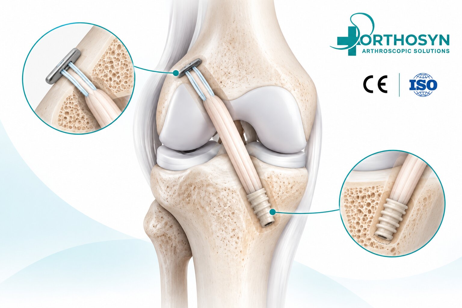
Hamstring tendon graft fixation in ACL reconstruction is a critical step in restoring knee stability after an anterior cruciate ligament (ACL) rupture. The procedure involves positioning a tendon graft within prepared bone tunnels or sockets and securing it to the femur and tibia using specialized orthopedic fixation devices.
Hamstring autografts, commonly harvested from the semitendinosus tendon and sometimes the gracilis tendon, are widely used in ACL reconstruction because they provide a biological graft capable of functioning as a ligament substitute after incorporation and remodeling.
However, successful ACL reconstruction depends not only on graft selection but also on accurate tunnel positioning, appropriate graft tension, fixation stability, biological healing, and postoperative rehabilitation.
Two important fixation principles are commonly used:
Suspensory fixation: A cortical button and loop construct secures the graft at the outer bone cortex.
Aperture fixation: An interference screw or another fixation device secures the graft closer to the entrance of the bone tunnel.
In many ACL reconstruction procedures, surgeons combine different fixation methods on the femoral and tibial sides.
The fundamental fixation concept is:
Hamstring Tendon Harvesting → Graft Preparation → Femoral and Tibial Tunnel Creation → Graft Passage → Femoral Fixation → Tibial Fixation → Graft Tensioning
Within the ORTHOSYN arthroscopy portfolio, Femoral Lift fixation systems, Fixed-Loop buttons, and Interference Screws represent different technologies used in orthopedic ligament reconstruction.
Understanding how these fixation systems work is important for orthopedic surgeons, sports medicine specialists, and medical device professionals.
A hamstring tendon graft is a biological tissue graft obtained from one or more tendons located along the posterior and medial regions of the thigh.
The most commonly used tendons include:
Semitendinosus tendon
Gracilis tendon
These tendons may be prepared individually or combined to create a graft of the required diameter and length.
Depending on the surgical technique, the graft may be folded into multiple strands.
Common configurations include:
Four-strand hamstring graft
Three-strand hamstring graft
Five- or six-strand graft constructs
All-inside ACL graft configurations
The selected graft configuration depends on available tendon length, graft diameter, fixation method, and surgical technique.
Hamstring tendons are used because they provide a strong biological tissue option for replacing a ruptured ACL.
Potential considerations include:
Availability of autologous tendon tissue
Ability to prepare different graft configurations
Avoidance of bone-block harvesting
Compatibility with several fixation systems
Established use in ACL reconstruction
However, hamstring grafts are not universally superior to patellar tendon or quadriceps tendon grafts.
Graft selection depends on patient characteristics, activity level, surgical objectives, and surgeon experience.
ACL reconstruction replaces a ruptured anterior cruciate ligament with a tendon graft.
The graft is positioned along a planned anatomical pathway between the femur and tibia.
The procedure generally involves:
Arthroscopic assessment of the knee
Hamstring tendon harvesting
Graft preparation and measurement
Femoral tunnel or socket preparation
Tibial tunnel or socket preparation
Graft passage
Femoral fixation
Tibial fixation
Graft tensioning and stability assessment
The objective is to establish a stable mechanical construct that supports the graft during biological incorporation.
Immediately after reconstruction, the tendon graft has not yet fully integrated into the surrounding bone.
The fixation devices must maintain graft position during the early healing period.
The fixation construct is exposed to forces generated by knee movement, rehabilitation, and daily activities.
Important fixation requirements include:
Resistance to graft slippage
Appropriate initial mechanical stability
Maintenance of graft tension
Compatibility with graft diameter
Accurate implant placement
Support for tendon-to-bone healing
The mechanical fixation system provides initial stability while biological incorporation develops.
However, fixation strength alone does not guarantee successful ACL reconstruction.
After harvesting, the tendon is cleaned and prepared according to the selected reconstruction technique.
Preparation may include:
Removal of residual muscle tissue
Tendon length measurement
Graft diameter measurement
Folding into multiple strands
Suturing of graft ends
Connection to a cortical suspensory fixation device
The graft must be compatible with the planned tunnel or socket dimensions.
For example, a four-strand hamstring graft may be connected to a femoral cortical button through a loop construct.
The opposite graft end may be prepared for tibial interference screw fixation or another compatible fixation method.
Femoral fixation secures the graft within the femoral tunnel or socket.
Common fixation methods include:
Fixed-loop cortical buttons
Adjustable-loop cortical buttons
Interference screws
Other compatible fixation devices
Suspensory cortical button fixation is frequently used with hamstring tendon grafts.
Cortical suspensory fixation uses a button positioned against the outer cortex of the femur.
A high-strength loop connects the button to the tendon graft.
The mechanical pathway is:
Femoral Cortex → Cortical Button → Suture Loop → Hamstring Tendon Graft
After the graft is introduced into the femoral socket, the button is deployed and positioned against the lateral femoral cortex.
The loop supports the graft and maintains its position within the prepared socket.
The surgeon must verify correct button deployment and positioning.
A fixed-loop femoral fixation system uses a cortical button connected to a loop of predetermined length.
The loop length is selected before implantation.
The graft is attached to the loop and positioned within the femoral socket.
The general sequence is:
Measure the femoral socket and planned graft position.
Select the appropriate fixed-loop length.
Attach the graft to the fixation loop.
Pass the button through the femoral tunnel.
Deploy the button outside the lateral femoral cortex.
Position the graft within the femoral socket.
Verify the fixation construct.
Fixed-loop systems require careful preoperative or intraoperative measurement because the loop length is not designed for subsequent shortening.
ORTHOSYN offers Fixed-Loop cortical fixation configurations with predetermined loop lengths.
The available range includes 15–45 mm, depending on the selected product configuration.
These systems represent a suspensory fixation option for applicable ligament reconstruction procedures.
An adjustable-loop femoral fixation system allows the surgeon to modify the effective loop length during implantation.
The button is positioned against the femoral cortex, and the graft can then be advanced into the socket by shortening the adjustable loop.
The basic sequence is:
Button Deployment → Loop Adjustment → Graft Advancement → Final Tensioning
This design provides intraoperative flexibility in graft positioning.
The surgeon generally:
Prepares the femoral socket.
Connects the graft to the adjustable-loop system.
Passes the cortical button through the femoral tunnel.
Deploys the button against the lateral femoral cortex.
Shortens the loop to advance the graft into the socket.
Adjusts graft positioning and tension according to the surgical technique.
Verifies button placement and fixation stability.
Some systems include additional locking or retensioning steps.
The specific procedure depends on the implant design and instructions for use.
ORTHOSYN provides adjustable-loop femoral fixation technologies within its ACL/PCL reconstruction portfolio.
Product Code: ORT-LS-1001
This system uses an adjustable suspensory fixation configuration.
It is designed around the principle of cortical button fixation with intraoperative loop adjustment.
Product Code: ORT-LS-3001
This system also utilizes an adjustable-loop cortical fixation concept.
The configuration provides another approach to graft positioning and loop adjustment.
Both product families represent suspensory fixation technologies.
Their exact deployment, adjustment, and locking sequences must follow the corresponding product instructions for use.
For product information, visit the ORTHOSYN official website.
Both systems use cortical suspensory fixation, but they differ in how loop length is established.
Characteristic | Fixed-Loop Fixation | Adjustable-Loop Fixation |
|---|---|---|
Loop length | Predetermined | Adjustable during implantation |
Graft positioning | Based on selected loop length | Can be adjusted after button deployment |
Femoral fixation | Cortical button | Cortical button |
Tunnel measurement | Important for loop selection | Important for graft positioning and adjustment |
Main consideration | Correct loop-length selection | Correct adjustment and locking |
ORTHOSYN examples | Fixed-Loop systems | Femoral Lift From Top / From Bottom |
Neither technology should be considered universally superior.
Biomechanical performance can vary between individual devices, and clinical outcomes depend on the complete reconstruction technique.
After femoral fixation, the graft must also be secured on the tibial side.
Tibial fixation methods include:
Interference screws
Cortical suspensory fixation
Screw-and-washer constructs
Other compatible fixation devices
Interference screw fixation is a commonly used method for securing soft tissue grafts within the tibial tunnel.
An interference screw is inserted into the bone tunnel alongside the tendon graft.
The screw creates mechanical compression between the graft and the tunnel wall.
The fixation principle is:
Tibial Bone Tunnel → Hamstring Graft → Interference Screw → Graft Compression
The objective is to limit graft movement and maintain fixation during the initial healing period.
The surgeon generally:
Positions the graft within the tibial tunnel.
Establishes the intended graft tension.
Positions the knee according to the selected fixation technique.
Introduces the interference screw alongside the graft.
Advances the screw to the planned depth.
Evaluates fixation stability.
Performs final graft tension and knee stability checks.
The screw diameter and length depend on tunnel dimensions, graft configuration, bone quality, and implant-specific recommendations.
Interference screws can be manufactured from different materials.
Common categories include:
Titanium
PEEK
Bioabsorbable polymers
Bioabsorbable polymer-ceramic composites
Each material has different mechanical, imaging, and degradation characteristics.
Titanium interference screws provide rigid metallic fixation.
They are nonabsorbable and remain in the bone unless removed for a specific clinical reason.
PEEK interference screws are nonabsorbable polymer implants.
They provide mechanical fixation without a metallic screw body.
Bioabsorbable screws are designed to undergo material degradation over time.
Their behavior depends on polymer composition, manufacturing characteristics, and the biological environment.
Degradation does not automatically imply complete replacement by native bone.
ORTHOSYN offers different interference screw technologies for applicable ligament reconstruction procedures.
Available diameters include:
7 mm
8 mm
9 mm
10 mm
Available lengths include:
25 mm
30 mm
ORTHOSYN also provides PEEK interference screw technology for compatible ligament fixation procedures.
The selected screw dimensions must match the planned tunnel and graft configuration.
The I-BONE portfolio includes:
PLA Bioabsorbable Interference Screws
Transparent polymer-based configurations.
PLDLLA + TCP Bioabsorbable Interference Screws
Polymer-ceramic composite configurations.
Available dimensions include:
Diameters: 7, 8, 9, and 10 mm
Lengths: 25 and 30 mm
The appropriate screw must be selected according to product indications, tunnel geometry, graft characteristics, and surgical requirements.
A common ACL reconstruction configuration combines femoral cortical suspensory fixation with tibial interference screw fixation.
Characteristic | Femoral Suspensory Fixation | Tibial Interference Screw Fixation |
|---|---|---|
Fixation location | Outer femoral cortex | Tibial bone tunnel |
Main device | Cortical button and loop | Interference screw |
Fixation mechanism | Suspensory support | Graft compression against bone |
Typical role | Femoral graft fixation | Tibial graft fixation |
Important consideration | Button deployment and loop position | Screw positioning and graft compression |
This combination uses two different mechanical principles to secure the same graft.
However, other femoral and tibial fixation combinations are also used in clinical practice.
Graft tensioning is an important part of ACL reconstruction.
Insufficient tension may contribute to residual laxity, while excessive tension may restrict knee movement or alter joint mechanics.
The surgeon evaluates graft tension according to the selected surgical technique.
Important considerations include:
Knee flexion angle
Graft positioning
Femoral fixation stability
Tibial fixation stability
Graft pretensioning
Cyclic knee movement
Final knee stability
The appropriate tensioning protocol depends on graft type, fixation system, and surgical technique.
There is no single universally appropriate tension value for every ACL reconstruction.
Graft-tunnel motion refers to movement of the graft within the prepared bone tunnel or socket.
Two commonly discussed biomechanical concepts are:
Longitudinal graft movement along the tunnel axis.
Transverse graft movement within the tunnel.
These phenomena are discussed in relation to fixation distance, graft-tunnel interaction, and tunnel enlargement.
However, the clinical significance depends on multiple factors, and the presence of suspensory fixation does not automatically mean that clinically important tunnel widening will occur.
The femoral and tibial tunnels establish the reconstructed graft pathway.
Incorrect tunnel positioning can alter graft tension and knee biomechanics.
Important considerations include:
Anatomical ACL footprint identification
Femoral tunnel orientation
Tibial tunnel positioning
Avoidance of graft impingement
Appropriate graft length within the socket
Compatibility with fixation devices
Even a mechanically strong fixation construct cannot fully compensate for substantially incorrect graft positioning.
Hamstring tendon grafts require biological incorporation into the surrounding bone.
This process involves cellular activity, tissue remodeling, and the development of an interface between the tendon and bone.
The simplified sequence is:
Initial Mechanical Fixation → Biological Incorporation → Graft Remodeling → Functional Adaptation
The process develops over months and is influenced by several factors.
Fixation devices provide mechanical support while biological healing progresses.
Important factors include:
Graft quality
Graft diameter
Tunnel positioning
Fixation stability
Patient age
Activity level
Biological healing capacity
Associated knee injuries
Rehabilitation compliance
Return-to-sport timing
No fixation device can eliminate all risks of graft failure.
Recovery varies according to the surgical technique, associated injuries, rehabilitation progress, and patient-specific factors.
Rehabilitation generally includes several stages.
The initial phase focuses on swelling management, restoration of knee extension, quadriceps activation, and safe mobility.
Strengthening exercises are gradually introduced for the quadriceps, hamstrings, and surrounding muscles.
Balance, coordination, and movement control are progressively developed.
Higher-demand activities are introduced after appropriate functional assessment.
Return to pivoting sports commonly requires at least 9 months, and some patients require longer.
Return-to-sport decisions should be based on objective functional criteria, psychological readiness, and clinical assessment rather than time alone.
Potential complications include:
Graft slippage
Fixation failure
Residual knee instability
Graft rupture
Tunnel widening
Knee stiffness
Infection
Persistent pain
Hardware-related complications
Delayed graft incorporation
The risk of complications depends on the patient, fixation method, surgical technique, and rehabilitation.
ORTHOSYN offers several fixation technologies relevant to ligament reconstruction procedures.
Adjustable-loop cortical suspensory fixation.
Reference: ORT-LS-1001
Adjustable-loop cortical suspensory fixation.
Reference: ORT-LS-3001
Cortical button fixation with predetermined loop lengths.
Metallic interference fixation technology.
Nonabsorbable polymer-based interference fixation.
PLA and PLDLLA + TCP screw configurations.
These product families support different principles of ACL graft fixation:
Femoral Cortical Fixation → Graft Positioning → Tibial Fixation → Mechanical Stability
The selected fixation construct must follow the specific device indications, compatibility requirements, and instructions for use.
For more information about available orthopedic fixation technologies, visit ORTHOSYN Medical.
Hamstring tendon graft fixation in ACL reconstruction involves securing a prepared tendon graft to the femur and tibia using compatible fixation devices.
Femoral fixation commonly uses cortical suspensory systems, including Fixed-Loop and Adjustable-Loop buttons.
Tibial fixation frequently uses interference screws manufactured from titanium, PEEK, or bioabsorbable materials.
The fundamental fixation principles are:
Femoral Cortex → Cortical Button → Suspensory Loop → Hamstring Graft
Tibial Tunnel → Hamstring Graft → Interference Screw → Graft Compression
Within the ORTHOSYN arthroscopy portfolio, Femoral Lift systems, Fixed-Loop devices, and Interference Screws represent different technologies for ligament reconstruction.
Successful ACL reconstruction depends on anatomical graft positioning, appropriate fixation stability, biological incorporation, and structured rehabilitation.