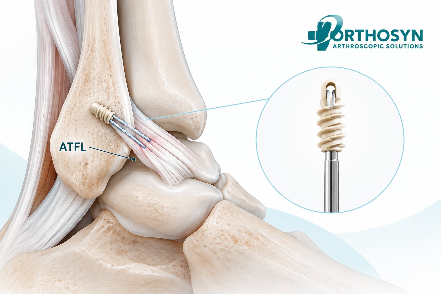
Anterior talofibular ligament (ATFL) repair is a surgical procedure used to restore the attachment and stability of one of the most frequently injured ligaments in the ankle. The ATFL plays an important role in maintaining lateral ankle stability, particularly during plantarflexion and inversion movements.
When the ATFL is damaged, patients may experience recurrent ankle sprains, pain, and chronic lateral ankle instability.
For selected patients with persistent mechanical instability despite appropriate conservative treatment, surgical repair may be considered.
One commonly performed technique is arthroscopic or open modified Broström repair, in which the damaged lateral ankle ligaments are repaired and reattached to their anatomical insertion using sutures and, in many techniques, suture anchors.
The fundamental fixation concept is:
ATFL Injury → Ligament Assessment → Fibular Footprint Preparation → Suture Anchor Placement → Ligament Reattachment → Healing
Within the ORTHOSYN arthroscopy portfolio, Titanium Suture Anchors, PEEK Suture Anchors, and Soft Anchorsrepresent different technologies for orthopedic soft tissue-to-bone fixation.
Understanding how suture anchors are used in ATFL repair is important for orthopedic surgeons, foot and ankle specialists, and sports medicine professionals.
The anterior talofibular ligament is part of the lateral ligament complex of the ankle.
This complex consists of three primary ligaments:
Anterior talofibular ligament (ATFL)
Calcaneofibular ligament (CFL)
Posterior talofibular ligament (PTFL)
The ATFL extends from the anterior region of the distal fibula to the talus.
Its primary functions include limiting excessive anterior translation of the talus and contributing to resistance against inversion, particularly when the ankle is plantarflexed.
Because of its anatomical orientation, the ATFL is especially vulnerable during inversion ankle sprains.
Ankle inversion injuries are common during sports and daily activities.
When the foot turns inward unexpectedly, the lateral ankle ligaments may experience excessive tensile forces.
The ATFL is often the first ligament affected.
More severe injuries may also involve the calcaneofibular ligament.
Repeated injuries can contribute to chronic lateral ankle instability.
ATFL injuries commonly result from excessive inversion or plantarflexion-inversion movements.
Contributing factors include:
Sports-related ankle sprains
Sudden changes in direction
Landing awkwardly after a jump
Walking or running on uneven surfaces
Recurrent inversion injuries
Previous ankle ligament damage
Inadequate recovery after an ankle sprain
ATFL injuries range from ligament stretching to complete rupture.
Not every ATFL tear requires surgery.
Many acute ankle sprains can be treated successfully with rehabilitation, bracing, and functional management.
Symptoms depend on the severity and chronicity of the injury.
Patients may experience:
Pain along the lateral ankle
Swelling
Tenderness near the distal fibula
Bruising after an acute injury
Difficulty bearing weight
Recurrent ankle sprains
A sensation of the ankle giving way
Reduced confidence during sports activities
Chronic instability may persist even after the initial swelling and pain have resolved.
Diagnosis generally involves clinical examination and imaging when appropriate.
The clinical assessment may include:
Anterior drawer test
Talar tilt test
Evaluation of lateral ankle tenderness
Assessment of ankle range of motion
Evaluation of balance and proprioception
Examination for associated ligament injuries
MRI can help evaluate ATFL integrity and associated soft tissue abnormalities.
Ultrasound may also be useful in selected cases.
Weight-bearing radiographs or other imaging may be used to investigate associated bone abnormalities or ankle alignment.
The surgeon must distinguish mechanical instability from functional instability and evaluate other possible causes of persistent symptoms.
Surgery may be considered when patients experience persistent symptomatic mechanical instability despite an adequate course of nonoperative treatment.
Conservative management may include:
Structured physiotherapy
Proprioceptive training
Peroneal muscle strengthening
Functional rehabilitation
Ankle bracing
Activity modification
Surgical treatment is not automatically indicated after an ATFL tear.
The decision depends on the degree of instability, functional limitations, ligament quality, associated injuries, and patient-specific factors.
Arthroscopic ATFL repair is a minimally invasive surgical approach used to restore lateral ankle ligament stability through small arthroscopic portals.
The surgeon evaluates the ankle joint, identifies the damaged ligament structures, and repairs the ATFL using an appropriate fixation technique.
Suture anchors may be inserted into the distal fibula to provide fixation points for the ligament and associated capsular tissue.
The basic repair principle is:
Distal Fibula → Suture Anchor → High-Strength Suture → ATFL Tissue
The sutures are used to approximate the ligament tissue to its anatomical fibular attachment.
In selected cases, the repair may also incorporate capsular reinforcement or treatment of associated calcaneofibular ligament pathology.
The modified Broström procedure is a commonly used surgical technique for treating chronic lateral ankle instability.
The original Broström concept involves direct repair of the injured lateral ankle ligaments.
The modified Broström-Gould technique additionally reinforces the repair using local tissue, traditionally including the inferior extensor retinaculum when appropriate.
Modern techniques may use suture anchors to facilitate anatomical ligament reattachment.
The procedure can be performed through open or arthroscopic approaches, depending on the clinical situation and surgical technique.
The principal objective is to restore lateral ankle stability while preserving physiological ankle movement.
Suture anchors create a mechanical connection between the distal fibula and the damaged ligament tissue.
The procedure generally involves several stages.
The surgeon examines the ankle joint and the lateral ligament structures.
Arthroscopic evaluation may include:
ATFL attachment region
Anterior ankle capsule
Talar cartilage
Tibial plafond
Synovial tissue
Associated intra-articular pathology
The surgeon evaluates the condition of the remaining ligament tissue and identifies associated injuries.
The damaged ATFL is assessed.
Important considerations include:
Tear location
Ligament tissue quality
Degree of attenuation
Remaining ligament attachment
Fibular footprint condition
Associated CFL injury
Mechanical instability
Direct repair is generally more suitable when sufficient ligament tissue remains available for secure reattachment.
Poor-quality or severely deficient tissue may require a different surgical strategy, including ligament reconstruction.
The anatomical fibular insertion of the ATFL is identified.
The surgeon prepares the relevant bone surface for ligament reattachment.
Preparation may involve removing residual soft tissue and creating an appropriate surface for fixation.
The objective is to position the repaired ligament near its native attachment.
Accurate anatomical positioning helps avoid excessive ligament tension or nonphysiological restriction of ankle movement.
One or more suture anchors are inserted into the distal fibula.
The number, diameter, and position of the anchors depend on:
Fibular bone dimensions
Bone quality
Ligament tissue quality
Repair configuration
Associated ligament injuries
Surgical technique
Device-specific indications
Because the distal fibula provides a relatively limited anatomical fixation area, anchor dimensions and placement require careful consideration.
The anchor establishes a fixation point for the sutures.
The sutures are passed through the remaining ATFL and, when appropriate, adjacent capsular tissue.
Different suture configurations may be used depending on the repair technique.
These may include:
Simple sutures
Mattress sutures
Modified mattress configurations
Knotless fixation techniques
The objective is to capture sufficient ligament tissue and create a stable repair construct.
Suture passage must account for tissue quality and the intended direction of ligament tension.
The ligament is approximated to its anatomical fibular insertion.
The sutures are secured using the selected technique.
In knotted repair systems, sutures are tied to maintain ligament approximation.
In knotless systems, the implant secures the sutures through its locking mechanism.
The surgeon aims to restore appropriate ligament tension without overtightening the lateral ankle structures.
After fixation, the surgeon evaluates the repair.
Assessment may include:
Ligament tension
Tissue approximation
Anchor stability
Ankle range of motion
Anterior translation
Inversion stability
Overall repair integrity
The ankle is assessed to confirm that the repair provides appropriate stability without excessive restriction.
Suture anchors allow ligament tissue to be secured directly to bone.
Their primary role is to maintain the repaired ligament in an appropriate position while biological healing develops.
Important mechanical considerations include:
Anchor fixation strength
Bone quality
Suture tensile strength
Ligament tissue capture
Suture configuration
Repair tension
Anatomical positioning
The simplified fixation pathway is:
Fibular Bone → Anchor Fixation → Suture Tension → Ligament Reattachment
However, mechanical fixation alone does not determine the clinical outcome.
Biological healing, tissue quality, associated injuries, and rehabilitation are also important.
Different suture anchor technologies are used in orthopedic soft tissue-to-bone fixation.
Common categories include:
Titanium suture anchors
PEEK suture anchors
All-suture or soft anchors
Bioabsorbable anchors
Each technology has different material and mechanical characteristics.
The selection depends on anatomical requirements, bone quality, fixation configuration, and the approved intended use of the device.
Titanium suture anchors provide rigid metallic fixation.
Their threaded or otherwise mechanically engaging structures are designed to obtain fixation within bone.
Potential considerations include:
Rigid implant construction
Mechanical engagement with bone
Availability of different diameters
Compatibility with high-strength sutures
Implant positioning within the distal fibula
Titanium anchors are used in various orthopedic ligament and tendon repair procedures.
Their suitability for ATFL repair depends on the specific anchor design, anatomical dimensions, and instructions for use.
ORTHOSYN offers titanium suture anchors in the following diameters:
Diameter | Product |
|---|---|
2.0 mm | Titanium Suture Anchor |
3.5 mm | Titanium Suture Anchor |
5.0 mm | Titanium Suture Anchor |
6.5 mm | Titanium Suture Anchor |
For ankle ligament repair, smaller anchor configurations may be considered when compatible with the patient's anatomy and the specific device indication.
The availability of a diameter does not automatically establish its suitability for ATFL repair.
PEEK, or polyetheretherketone, is a nonabsorbable polymer used in orthopedic implants.
PEEK suture anchors provide rigid fixation without a metallic anchor body.
Their performance depends on anchor geometry, bone engagement, insertion technique, and bone quality.
The ORTHOSYN portfolio includes PEEK suture anchors in:
4.5 mm
5.5 mm
6.5 mm
ORTHOSYN also offers knotless anchor configurations:
PEEK + PEEK Knotless Anchors
3.5 mm
5.0 mm
PEEK + Titanium Knotless Anchors
3.5 mm
5.0 mm
Because the distal fibula has limited bone dimensions, anchor size and configuration must be evaluated carefully.
Larger anchors should not automatically be assumed appropriate for ATFL repair.
The selected device must be compatible with the intended anatomical application.
Soft anchors, also known as all-suture anchors, use high-strength textile structures rather than a conventional rigid anchor body.
During insertion, the textile component is deployed within the bone to create a fixation mechanism.
Depending on the design, soft anchors may require smaller bone openings than certain rigid anchors.
This characteristic can be relevant in anatomical regions with limited bone stock.
However, fixation behavior depends on:
Bone quality
Deployment technique
Anchor design
Insertion depth
Loading conditions
Suture configuration
ORTHOSYN Soft Anchors use UHMWPE-based textile technology.
Available diameters include:
1.3 mm
1.5 mm
1.9 mm
3.0 mm
These products represent different textile-based fixation options within the ORTHOSYN arthroscopy portfolio.
Their suitability for ATFL repair must be confirmed using the specific product indications and instructions for use.
Characteristic | Titanium Anchor | PEEK Anchor | Soft Anchor |
|---|---|---|---|
Main material | Titanium | PEEK polymer | UHMWPE-based textile |
Structure | Rigid | Rigid | Flexible textile |
Fixation principle | Mechanical bone engagement | Mechanical bone engagement | Textile deployment within bone |
ORTHOSYN sizes | 2.0–6.5 mm | 4.5–6.5 mm | 1.3–3.0 mm |
Main anatomical consideration | Bone dimensions and placement | Bone dimensions and placement | Bone quality and deployment |
ATFL application | Product-specific | Product-specific | Product-specific |
No single anchor material is universally superior for ATFL repair.
The appropriate choice depends on the patient's anatomy, surgical technique, fixation requirements, and device documentation.
Suture anchors may be used in knotted or knotless repair configurations.
In a knotted repair, sutures are passed through the ligament tissue and tied to maintain approximation.
The surgeon controls the fixation through suture placement, tensioning, and knot tying.
Knotless fixation systems secure sutures using an implant-based locking mechanism.
This eliminates the need for conventional knot tying at the final fixation point.
Potential technical considerations include:
Suture management
Ligament tensioning
Working space
Anchor positioning
Tissue compression
Neither approach should be considered universally superior.
Clinical performance depends on the complete repair construct and surgical technique.
The ATFL contributes to controlling anterior talar translation and inversion-related instability.
Restoring the ligament near its native anatomical attachments helps preserve its intended mechanical function.
A nonanatomical or excessively tensioned repair may alter ankle motion.
The objective is:
Anatomical Reattachment → Mechanical Stability → Ligament Healing → Functional Recovery
However, anatomical fixation does not guarantee complete recovery.
The outcome also depends on tissue quality, associated injuries, and rehabilitation.
ATFL repair and reconstruction are different surgical strategies.
Repair uses the patient's remaining ligament tissue.
The damaged ligament is reattached or reinforced using sutures and fixation devices.
Reconstruction uses a graft or another tissue substitute to recreate the ligament when direct repair is not appropriate.
This may be considered in selected cases involving:
Severely deficient ligament tissue
Failed previous repair
Complex chronic instability
Poor-quality residual ligament structures
The choice between repair and reconstruction requires individual clinical assessment.
Yes.
The ATFL and calcaneofibular ligament are both components of the lateral ankle ligament complex.
Some patients have injuries involving both ligaments.
Depending on the injury pattern, the surgeon may repair or reconstruct both structures.
The fixation strategy depends on the location and quality of the injured tissues.
Not every ATFL injury requires CFL repair.
High-strength sutures connect the ligament tissue to the bone fixation device.
Their function is to maintain tissue approximation during the initial healing period.
Important characteristics include:
Tensile strength
Abrasion resistance
Handling properties
Knot security, where applicable
Compatibility with anchor systems
Resistance to tissue cut-through
UHMWPE-based sutures are used in different orthopedic soft tissue repair procedures.
However, successful fixation depends on the entire repair construct rather than suture strength alone.
Recovery varies depending on the surgical technique, associated procedures, tissue quality, and patient-specific factors.
Rehabilitation generally progresses through several phases.
The repaired ligament is protected during initial healing.
Immobilization or a protective boot may be used according to the surgeon's protocol.
Ankle movement is gradually introduced within prescribed limits.
Inversion stress may be restricted during early rehabilitation.
Strengthening and proprioceptive exercises are introduced as healing progresses.
Balance, coordination, and sport-specific activities are gradually restored.
Return to sports often takes several months, depending on the procedure and clinical progress.
The rehabilitation protocol must be individualized by the treating surgeon.
Ligament healing depends on several mechanical and biological factors.
These include:
Ligament tissue quality
Chronicity of the injury
Anatomical repair position
Fixation stability
Associated CFL injury
Patient activity level
Previous ankle injuries
Rehabilitation compliance
General health and healing capacity
Stable fixation supports the healing process, but it cannot eliminate all biological risks.
Potential complications may include:
Recurrent ankle instability
Persistent pain
Ankle stiffness
Wound complications
Infection
Nerve irritation or injury
Anchor-related complications
Incomplete ligament healing
Restricted ankle motion
Failure of the repair construct
The likelihood of complications depends on the patient, injury characteristics, and surgical technique.
ORTHOSYN offers several suture anchor technologies for applicable orthopedic soft tissue-to-bone fixation procedures.
The portfolio includes:
ORTHOSYN Titanium Suture Anchors
Rigid metallic fixation technology in multiple diameters.
ORTHOSYN PEEK Suture Anchors
Nonabsorbable polymer-based fixation technology.
ORTHOSYN Soft Anchors
UHMWPE-based textile fixation technology.
ORTHOSYN Knotless Anchors
PEEK + PEEK and PEEK + Titanium fixation configurations.
ORTHOSYN Enhanced Sutures
High-strength UHMWPE-based braided sutures.
These technologies represent different approaches to the general principles of orthopedic ligament fixation:
Bone Preparation → Anchor Placement → Suture Passage → Ligament Reattachment → Mechanical Stabilization
Their use in ATFL repair must follow the specific product indications, instructions for use, and supporting technical documentation.
Explore the ORTHOSYN Suture Anchors portfolio for more information about available anchor technologies.
Suture anchors are used in ATFL repair to secure damaged lateral ankle ligament tissue to its anatomical attachment on the distal fibula.
The procedure generally involves ligament assessment, fibular footprint preparation, anchor placement, suture passage, ligament reattachment, and stability evaluation.
The fundamental fixation principle is:
Distal Fibula → Suture Anchor → High-Strength Suture → ATFL Reattachment
Different anchor technologies, including titanium, PEEK, and all-suture anchors, provide distinct fixation characteristics.
Within the ORTHOSYN arthroscopy portfolio, these technologies are represented by multiple suture anchor configurations and UHMWPE-based sutures.
Successful ATFL repair depends on appropriate patient selection, anatomical fixation, ligament tissue quality, biological healing, and postoperative rehabilitation.