


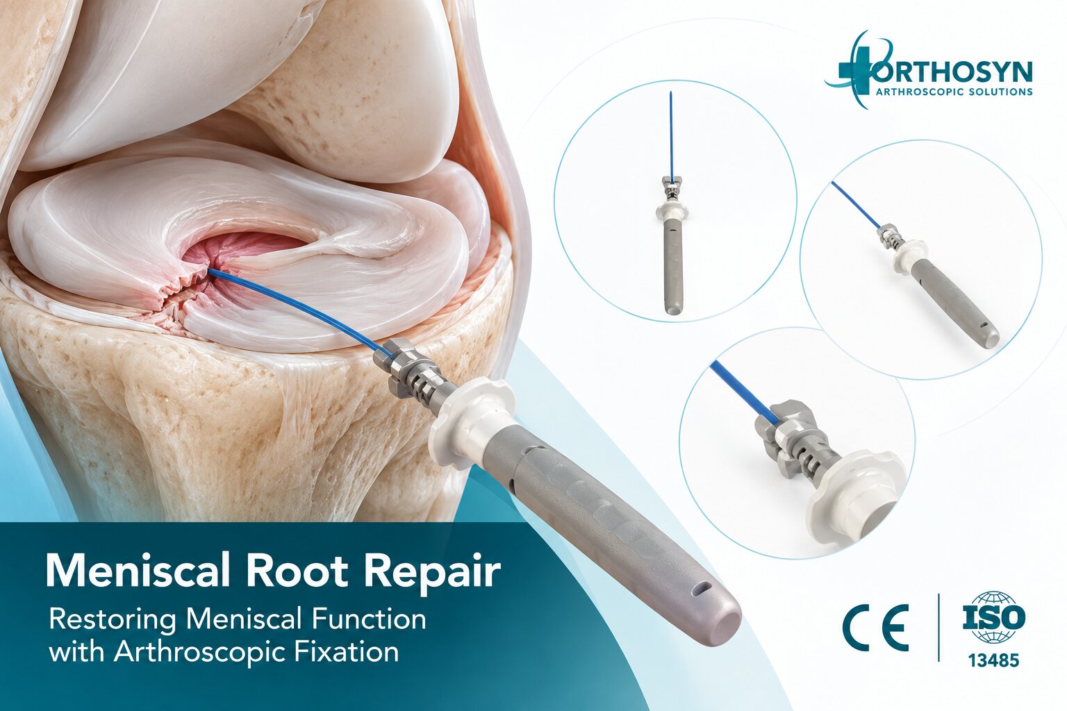
Meniscal root repair is a surgical procedure used to restore the attachment of a torn meniscal root to its anatomical insertion on the tibia. The meniscal roots are essential for maintaining knee stability, distributing joint loads, and preserving the shock-absorbing function of the meniscus.
When a meniscal root is torn, the meniscus may lose its ability to convert compressive forces into circumferential tension, commonly known as hoop stress. This can lead to meniscal extrusion, increased contact pressure on the articular cartilage, and accelerated joint degeneration.
Arthroscopic meniscal root repair aims to restore the meniscus-to-bone attachment through specialized suturing and fixation techniques.
Two important fixation approaches include:
Transtibial pullout repair: Sutures are passed through the meniscal root and secured through one or more tibial bone tunnels.
Suture anchor repair: The torn meniscal root is reattached near its anatomical footprint using a suitable anchor-based fixation construct.
Understanding these techniques is important for orthopedic surgeons and sports medicine professionals involved in arthroscopic knee surgery.
Within the ORTHOSYN arthroscopy portfolio, meniscal repair technologies are part of a broader range of solutions for treating selected meniscal injuries.
The menisci are fibrocartilaginous structures located between the femur and tibia.
Each knee contains two menisci:
Medial meniscus
Lateral meniscus
Each meniscus has anterior and posterior root attachments that secure it to the tibial plateau.
These attachments allow the meniscus to remain positioned within the joint during movement and loading.
The meniscal roots play an essential role in transmitting forces between the meniscus and the underlying bone.
During weight-bearing activities, the meniscus experiences compressive forces.
Its circumferential collagen fibers help convert these forces into hoop tension.
The meniscal root attachments provide the anchoring points necessary for this mechanism.
The simplified biomechanical pathway is:
Joint Compression → Meniscal Hoop Stress → Root Attachment → Load Distribution
When a root attachment is disrupted, the meniscus may no longer distribute loads effectively.
This can increase stress on the articular cartilage.
A meniscal root tear is an injury involving the attachment of the meniscus to the tibia or the tissue immediately adjacent to that attachment.
Root tears may involve complete detachment or a radial tear close to the root insertion.
These injuries can significantly affect meniscal biomechanics.
Meniscal root tears are commonly categorized as:
Posterior medial meniscal root tears are frequently associated with degenerative changes, although traumatic injuries can also occur.
They may be accompanied by meniscal extrusion and cartilage degeneration.
Posterior lateral meniscal root tears are often associated with traumatic knee injuries, particularly anterior cruciate ligament (ACL) tears.
These injuries may affect rotational stability and lateral compartment biomechanics.
Anterior root tears are less frequently discussed but can also compromise meniscal attachment and function.
The clinical significance depends on the location, tear configuration, and associated knee pathology.
Meniscal root tears may develop through traumatic or degenerative mechanisms.
Common contributing factors include:
Sports-related knee injuries
Sudden twisting movements
Deep knee flexion under load
ACL injuries
Age-related meniscal degeneration
Repetitive mechanical stress
Increased body weight
Degenerative cartilage changes
The underlying mechanism may differ between medial and lateral meniscal root injuries.
Not every root tear is caused by a single traumatic event.
Symptoms vary depending on the tear location and associated joint conditions.
Patients may experience:
Knee pain
Joint-line tenderness
Swelling
Pain during squatting
Difficulty with deep knee flexion
Reduced functional capacity
A popping sensation at the time of injury
Discomfort during weight-bearing activities
Some degenerative meniscal root tears develop without a clearly identifiable injury.
Clinical assessment and imaging are important for diagnosis.
Diagnosis typically involves clinical examination and magnetic resonance imaging (MRI).
MRI may demonstrate:
Meniscal root discontinuity
Radial tears near the root attachment
Meniscal extrusion
The characteristic ghost meniscus sign
Associated cartilage damage
Bone marrow abnormalities
Concomitant ligament injuries
Arthroscopy can provide direct visualization and probing of the meniscal root when surgical treatment is undertaken.
The surgeon evaluates whether the root is detached, whether the tissue is repairable, and whether associated knee abnormalities require treatment.
The meniscal root is essential for maintaining the meniscus in its functional position.
When the root is detached, the meniscus may lose its ability to transmit hoop stresses.
Biomechanical studies have shown that complete posterior medial meniscal root tears can substantially increase tibiofemoral contact pressures.
In certain experimental conditions, the biomechanical consequences may resemble those of extensive meniscal tissue loss.
However, the degree of functional impairment depends on the specific tear and joint conditions.
The primary objectives of meniscal root repair are to:
Restore the anatomical root attachment
Improve meniscal stability
Re-establish load transmission
Reduce abnormal meniscal displacement
Support preservation of joint function
Repair does not guarantee prevention or reversal of osteoarthritis.
Arthroscopic meniscal root repair is a minimally invasive procedure that uses an arthroscope and specialized instruments to restore the attachment of the meniscal root to the tibia.
The surgeon identifies the torn root, prepares its anatomical attachment site, passes sutures through the meniscal tissue, and secures the repair.
The fixation must maintain contact between the meniscal root and the prepared bone surface during biological healing.
The fundamental repair sequence is:
Root Tear Identification → Footprint Preparation → Suture Passage → Root Reduction → Fixation → Meniscus-to-Bone Healing
The specific technique depends on the root involved, tear pattern, bone anatomy, and surgeon preference.
The procedure generally involves several stages.
The surgeon introduces the arthroscope and examines the knee joint.
Important structures include:
Medial and lateral menisci
Anterior and posterior meniscal roots
Tibial plateau
Femoral cartilage
ACL and PCL
Surrounding joint structures
The surgeon identifies the root tear and evaluates associated injuries.
The torn meniscal root is evaluated using arthroscopic instruments.
Important considerations include:
Tear configuration
Tissue quality
Root displacement
Meniscal extrusion
Mobility of the torn tissue
Anatomical footprint location
The surgeon determines whether the root can be reduced to its anatomical attachment without excessive tension.
The original root footprint on the tibia is identified.
The attachment area may be prepared to expose an appropriate bone surface for healing.
The objective is to create a suitable interface between the meniscal tissue and the tibial bone.
Accurate anatomical positioning is important because nonanatomical fixation may alter meniscal biomechanics.
High-strength sutures are passed through the torn root tissue.
Different suture configurations may be used, including:
Simple sutures
Mattress sutures
Cinch sutures
Luggage-tag configurations
Other root-specific suture patterns
The selected configuration should provide adequate tissue capture and minimize the risk of suture cut-through.
The sutures are tensioned to bring the meniscal root toward its anatomical footprint.
The surgeon evaluates tissue positioning and tension.
The objective is to restore contact between the root and the tibial attachment site.
The meniscal root is secured using the selected fixation technique.
Common approaches include transtibial pullout fixation and suture anchor fixation.
The fixation construct should maintain root positioning while healing develops.
The surgeon evaluates:
Root position
Suture tension
Fixation stability
Tissue approximation
Meniscal mobility
Associated joint pathology
The repair is assessed before the procedure is completed.
Transtibial pullout repair is a widely used technique for arthroscopic meniscal root fixation.
The procedure involves passing sutures through the torn meniscal root and directing them through one or more bone tunnels created in the tibia.
The sutures are then secured on the anterior tibial cortex using an appropriate fixation method.
The mechanical pathway is:
Meniscal Root → High-Strength Sutures → Tibial Tunnel → Cortical Fixation
This creates a connection between the meniscal root and the tibial bone.
The surgeon generally:
Identifies the anatomical root footprint.
Prepares the tibial attachment site.
Passes sutures through the torn root.
Creates a tibial tunnel or tunnels.
Retrieves the sutures through the tunnel.
Reduces the root to its footprint.
Secures the sutures at the tibial cortex.
Assesses the stability of the repair.
The technique may use cortical buttons, fixation anchors, or other suitable devices depending on the surgical system.
The exact fixation method must be compatible with the selected instruments and implants.
Suture anchor repair uses an implant positioned near the meniscal root footprint to secure the torn tissue to bone.
Rather than relying on a long transtibial pullout construct, this approach creates fixation closer to the root insertion.
The simplified concept is:
Tibial Root Footprint → Suture Anchor → High-Strength Sutures → Meniscal Root
The technique may be considered in selected anatomical situations.
However, arthroscopic access, anchor placement, bone quality, and available working space influence its feasibility.
Not every suture anchor is designed or approved for meniscal root repair.
The surgeon must verify the specific implant's intended purpose and instructions for use.
Both techniques aim to restore the meniscal root attachment, but their fixation configurations differ.
Characteristic | Transtibial Pullout Repair | Suture Anchor Repair |
|---|---|---|
Fixation location | Tibial cortex through bone tunnel | Near the meniscal root footprint |
Bone preparation | Tibial tunnel drilling | Anchor socket or insertion site |
Suture pathway | Root through tibial tunnel | Root to local anchor |
Fixation principle | Transosseous suture fixation | Anchor-based fixation |
Main consideration | Tunnel positioning and suture construct | Anchor positioning and access |
Clinical selection | Patient- and technique-specific | Patient- and technique-specific |
Neither technique should be considered universally superior.
The optimal approach depends on the tear, anatomy, surgeon experience, and device-specific characteristics.
Sutures form the mechanical connection between the meniscal root and the fixation construct.
Their function is to maintain root approximation during the early healing period.
Important suture characteristics include:
Tensile strength
Abrasion resistance
Knot security, where applicable
Compatibility with the fixation device
Handling properties
Resistance to tissue cut-through
UHMWPE-based sutures are used in various orthopedic soft tissue repair applications.
However, successful fixation depends on the entire construct, including tissue quality, suture configuration, bone fixation, and surgical technique.
The meniscal root attachment has a specific anatomical location.
Restoring the root near its original footprint is important for re-establishing normal meniscal function.
A root repaired in a substantially displaced position may not reproduce the intended biomechanical relationship.
The objective is:
Anatomical Root Position → Stable Fixation → Biological Healing → Improved Meniscal Function
Nevertheless, successful healing and functional recovery cannot be guaranteed solely through anatomical positioning.
Meniscal extrusion refers to displacement of the meniscus beyond the edge of the tibial plateau.
It may occur when the meniscal root attachment is disrupted.
Extrusion can reduce the effective contact area through which the meniscus distributes loads.
Meniscal root repair aims to restore the root attachment, but extrusion may persist in some patients.
Additional procedures, including meniscal centralization techniques, may be considered in selected cases.
These are separate surgical decisions requiring individual evaluation.
Yes.
Lateral meniscal posterior root tears can occur in association with ACL injuries.
When both injuries are present, the surgeon may perform meniscal root repair and ACL reconstruction during the same procedure.
However, surgical planning must account for:
Tibial tunnel positioning
Potential tunnel convergence
Graft fixation
Root fixation
Associated meniscal damage
Rehabilitation requirements
The surgeon must ensure that the fixation constructs do not interfere with each other.
Standard meniscal repair generally addresses tears within the body or horns of the meniscus.
Meniscal root repair specifically restores the attachment between the meniscus and the tibia.
Characteristic | Standard Meniscal Repair | Meniscal Root Repair |
|---|---|---|
Primary injury | Tear within meniscal tissue | Root detachment or tear near insertion |
Main objective | Approximate torn meniscal tissue | Restore meniscus-to-bone attachment |
Common techniques | All-inside, inside-out, outside-in | Transtibial pullout, root-specific anchor fixation |
Fixation target | Meniscal tissue | Meniscal root and tibial footprint |
Bone tunnel | Usually not required | Common in pullout repair |
These differences are important when selecting instrumentation and fixation systems.
All-inside meniscal repair systems are designed to approximate selected tears within the meniscal tissue using implants and sutures.
They can reduce the need for additional surgical incisions in appropriate meniscal repair procedures.
However, conventional all-inside meniscal repair systems and meniscal root fixation systems are not interchangeable.
Root repair requires a fixation construct capable of restoring the meniscus-to-bone attachment.
An all-inside repair implant should not automatically be considered suitable for root fixation.
Its intended purpose and instructions for use must specifically support the planned procedure.
ORTHOSYN provides meniscal repair technologies for selected arthroscopic knee procedures.
The portfolio includes the ORTHOSYN VersaFix Meniscal Repair System, which incorporates:
Two PEEK fixation implants
High-strength UHMWPE suture
Adjustable depth-control mechanism
An all-inside meniscal repair configuration
VersaFix is associated with the repair of selected meniscal tissue tears using an all-inside fixation approach.
It is important to distinguish this from meniscal root fixation.
The presence of PEEK implants and high-strength sutures does not automatically establish an indication for transtibial or anchor-based meniscal root repair.
Product selection must follow the specific approved indications and instructions for use.
For detailed product information, visit the official ORTHOSYN Meniscal Repair Systems category.
After meniscal root fixation, the surgeon assesses whether the root remains appropriately positioned against its attachment site.
Important considerations include:
Suture tension
Root reduction
Fixation stability
Tissue capture
Implant positioning
Tibial tunnel configuration, when applicable
Meniscal mobility
The construct should maintain sufficient stability during the initial healing period.
However, mechanical stability alone does not guarantee biological healing.
Meniscal root healing depends on several biological and mechanical factors.
These include:
Tear chronicity
Tissue quality
Anatomical reduction
Fixation stability
Patient age
Cartilage condition
Meniscal extrusion
Limb alignment
Body weight
Rehabilitation compliance
Advanced joint degeneration or substantial malalignment may affect treatment selection and expected outcomes.
The decision to repair a meniscal root should be individualized.
Recovery varies according to the tear, fixation technique, associated procedures, and surgeon's protocol.
Rehabilitation generally includes several phases.
Weight-bearing and knee flexion may be restricted to protect the repair.
Knee movement is gradually introduced within prescribed limits.
Muscle strengthening and functional exercises are advanced according to healing progress.
Higher-demand activities and sports are resumed gradually after appropriate clinical assessment.
Recovery often requires several months.
Return to sport may take approximately six months or longer, depending on the procedure and patient-specific factors.
The rehabilitation plan must be determined by the treating surgeon.
Potential complications include:
Failure of root healing
Recurrent meniscal tear
Persistent meniscal extrusion
Suture cut-through
Fixation failure
Knee stiffness
Infection
Persistent pain
Progression of cartilage degeneration
The likelihood of complications depends on patient characteristics, tissue quality, joint condition, and surgical technique.
Meniscal root repair aims to preserve the meniscus and restore its attachment, while meniscectomy removes damaged meniscal tissue.
In appropriately selected patients, root repair may offer advantages for meniscal preservation and joint biomechanics.
Clinical studies have reported favorable outcomes for root repair compared with meniscectomy in certain patient groups.
However, not all root tears are repairable, and the evidence does not establish that repair is superior for every patient.
Advanced osteoarthritis, substantial malalignment, poor tissue quality, or other factors may influence treatment selection.
ORTHOSYN focuses on providing specialized technologies for orthopedic arthroscopy.
Its meniscal repair portfolio includes solutions intended for selected meniscal tissue repair procedures.
The ORTHOSYN VersaFix Meniscal Repair System combines PEEK implants, UHMWPE suture technology, and adjustable depth control in an all-inside configuration.
These technologies are relevant to broader meniscal preservation principles, including tissue approximation and stable fixation.
For root-specific procedures, the selected fixation system must be verified as appropriate for meniscus-to-bone attachment repair.
Explore the complete ORTHOSYN Meniscal Repair Systems product portfolio.
Meniscal root repair is an arthroscopic surgical procedure intended to restore the attachment of a torn meniscal root to the tibia.
The meniscal roots are essential for maintaining hoop stress transmission, distributing joint loads, and preserving meniscal function.
Two established fixation approaches are:
Transtibial Pullout Repair → Meniscal Sutures → Tibial Tunnel → Cortical Fixation
Suture Anchor Repair → Local Bone Anchor → Suture Fixation → Root Reattachment
Both techniques aim to maintain anatomical root positioning during biological healing.
Within the ORTHOSYN arthroscopy portfolio, meniscal repair technologies support selected arthroscopic meniscal preservation procedures.
The appropriate surgical technique and fixation device must be selected according to the patient's anatomy, tear characteristics, product indications, and the surgeon's clinical judgment.