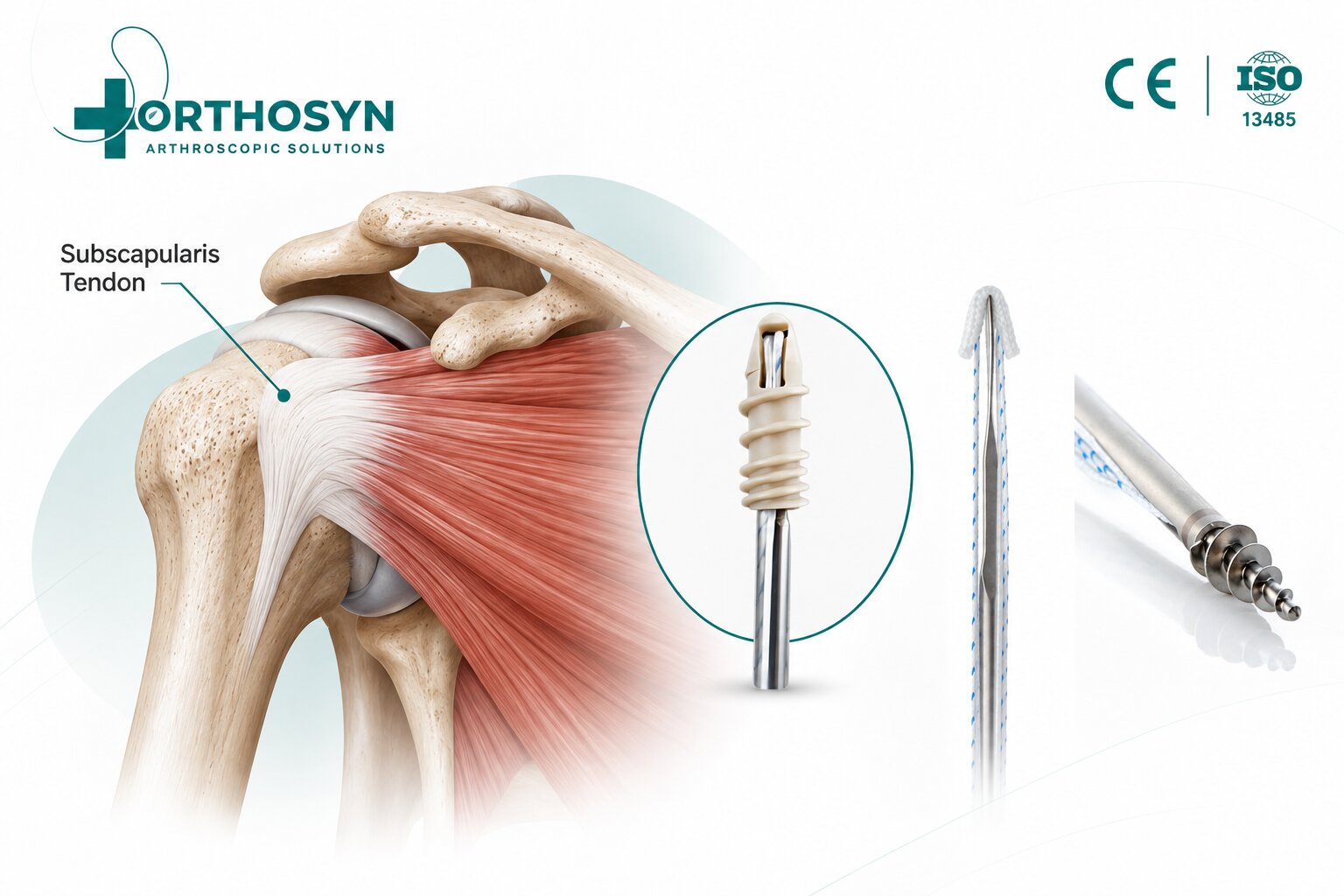
Arthroscopic subscapularis tendon repair is a surgical technique used to restore the attachment of a torn subscapularis tendon to the lesser tuberosity of the humerus. As an important component of the rotator cuff, the subscapularis contributes to shoulder stability, internal rotation, and coordinated glenohumeral movement.
Subscapularis tendon tears may occur following traumatic injuries, chronic degeneration, or in association with other rotator cuff injuries.
When surgical treatment is indicated, arthroscopic repair allows the surgeon to visualize the damaged tendon, prepare its anatomical attachment site, and secure the tendon to bone using suture anchors.
The fundamental fixation concept is:
Subscapularis Tendon Tear → Arthroscopic Evaluation → Tendon Mobilization → Suture Anchor Fixation → Tendon-to-Bone Healing
Within the ORTHOSYN arthroscopy portfolio, PEEK Suture Anchors, Titanium Suture Anchors, and Soft Anchorsrepresent different technologies for orthopedic soft tissue-to-bone fixation.
Understanding the anatomy, surgical steps, and fixation principles of subscapularis repair is important for orthopedic surgeons and sports medicine professionals.
The subscapularis is one of the four muscles forming the rotator cuff.
The rotator cuff consists of:
Supraspinatus
Infraspinatus
Teres minor
Subscapularis
The subscapularis muscle originates from the anterior surface of the scapula and its tendon inserts primarily onto the lesser tuberosity of the humerus.
Its main functions include internal rotation of the shoulder and stabilization of the humeral head within the glenoid.
The subscapularis also contributes to the anterior force couple of the shoulder, working with other rotator cuff muscles to maintain balanced joint movement.
Because of its anatomical position, subscapularis injuries may affect both shoulder strength and stability.
Subscapularis tendon tears can result from acute trauma or progressive tendon degeneration.
Common causes and contributing factors include:
Traumatic shoulder injuries
Falls onto an outstretched arm
Forced external rotation
Shoulder dislocation
Age-related tendon degeneration
Repetitive mechanical loading
Chronic rotator cuff disease
Associated supraspinatus tendon tears
Some tears involve only the upper portion of the subscapularis tendon, while others extend across a larger portion of its insertion.
Subscapularis tears may also be associated with abnormalities of the long head of the biceps tendon and its stabilizing pulley structures.
Symptoms vary according to the size, location, and chronicity of the injury.
Patients may experience:
Anterior shoulder pain
Weakness during internal rotation
Difficulty reaching behind the back
Pain during lifting activities
Reduced shoulder function
Discomfort during overhead movement
Associated biceps tendon symptoms
Some patients may experience weakness without substantial pain.
Clinical examination and imaging help determine whether the subscapularis tendon is injured.
Diagnosis generally involves a combination of clinical examination and imaging.
Physical examination may include:
Lift-off test
Belly-press test
Bear-hug test
Internal rotation strength assessment
Evaluation of shoulder range of motion
Assessment of associated rotator cuff pathology
MRI is commonly used to evaluate tendon integrity, tear dimensions, retraction, and muscle quality.
Ultrasound may also be useful when performed by an experienced examiner.
The surgeon evaluates whether the tear is partial-thickness or full-thickness and determines the extent of tendon involvement.
Subscapularis tendon tears may be classified according to their anatomical extent and tissue characteristics.
Common patterns include:
Only part of the tendon thickness is affected.
These injuries may involve the articular surface or a limited portion of the tendon insertion.
The superior portion of the subscapularis tendon is detached or damaged.
These tears are particularly relevant because the upper subscapularis is closely associated with the biceps pulley.
The tear extends through the entire tendon thickness.
The extent of the detached footprint may vary.
The tendon has moved away from its anatomical insertion.
Mobilization may be necessary before repair.
The surgical approach depends on tear configuration, tissue quality, tendon mobility, and associated shoulder pathology.
Arthroscopic subscapularis repair is a minimally invasive surgical procedure performed using an arthroscope and specialized instruments introduced through small portals.
The objective is to restore the attachment of the damaged tendon to the lesser tuberosity.
Suture anchors are commonly used to provide fixation points within the humerus.
The sutures are passed through the tendon and secured to approximate the tissue to its anatomical footprint.
The simplified repair concept is:
Lesser Tuberosity → Suture Anchor → High-Strength Suture → Subscapularis Tendon
Arthroscopic visualization allows the surgeon to assess the tear, evaluate associated structures, and perform the repair according to the selected technique.
The procedure generally involves several stages.
The surgeon introduces the arthroscope into the shoulder joint and evaluates the subscapularis tendon.
Important structures include:
Subscapularis tendon
Lesser tuberosity
Long head of the biceps tendon
Biceps pulley
Supraspinatus tendon
Glenohumeral cartilage
Surrounding capsuloligamentous structures
The surgeon determines the extent of the tear and identifies associated pathology.
The biceps tendon is particularly important because instability or damage may accompany superior subscapularis tears.
The torn portion of the subscapularis tendon is identified.
The surgeon assesses:
Tear location
Tendon retraction
Tissue quality
Remaining tendon attachment
Footprint involvement
Tendon mobility
A probe may be used to evaluate the tendon and confirm the tear configuration.
The repair strategy is selected according to these findings.
Retracted subscapularis tears may require mobilization before the tendon can be returned to its anatomical insertion.
The surgeon may release adhesions or surrounding scar tissue when necessary.
The objective is to achieve adequate tendon mobility without excessive tension.
A tension-free or appropriately tensioned repair is important because excessive tension may compromise fixation or healing.
The lesser tuberosity is the primary insertion site of the subscapularis tendon.
The surgeon prepares the relevant bone surface to support tendon reattachment.
Preparation may involve removing residual soft tissue and creating an appropriate fixation surface while preserving necessary bone stock.
The objective is to establish a suitable tendon-to-bone healing environment.
One or more suture anchors are inserted into the lesser tuberosity.
The number and position of anchors depend on:
Tear size
Footprint dimensions
Tendon quality
Bone quality
Repair configuration
Surgical technique
The anchors establish fixation points for the sutures used to secure the tendon.
The basic fixation pathway is:
Humeral Bone → Suture Anchor → Suture → Subscapularis Tendon
The sutures connected to the anchors are passed through the subscapularis tendon.
Depending on the technique, the surgeon may use:
Simple suture configurations
Mattress sutures
Modified mattress configurations
Knotless fixation techniques
Other tendon-specific suture patterns
The goal is to obtain secure tendon capture and distribute fixation forces appropriately.
Suture placement should account for tendon thickness, tissue quality, and the direction of the repair.
The tendon is positioned against the prepared lesser tuberosity footprint.
The sutures are secured using the selected fixation method.
In a knotted repair, sutures are tied to maintain tendon approximation.
In a knotless repair, the fixation system secures the sutures without requiring conventional knot tying at the final fixation point.
The objective is to restore tendon contact with its anatomical insertion while avoiding excessive tension.
After fixation, the surgeon evaluates the completed repair.
Assessment may include:
Tendon position
Footprint coverage
Suture tension
Anchor stability
Tendon mobility
Repair integrity during controlled shoulder movement
Associated shoulder pathology may also be addressed according to the surgical plan.
Suture anchors provide a mechanical connection between the humerus and the torn tendon.
Their primary role is to maintain tendon approximation to bone during the healing process.
Important fixation considerations include:
Anchor stability
Bone quality
Suture strength
Tendon capture
Footprint restoration
Load distribution
Repair tension
The mechanical construct must maintain sufficient stability while biological tendon-to-bone healing develops.
However, fixation strength alone does not determine clinical success.
Tendon quality, patient characteristics, rehabilitation, and biological healing also influence outcomes.
Subscapularis repair can involve different anchor configurations.
Single-row repair generally uses one row of anchors to secure the tendon to the lesser tuberosity.
This approach may be considered for selected smaller or less extensive tears.
The configuration depends on the tear pattern and surgical technique.
Double-row repair uses a broader fixation configuration involving medial and lateral fixation points.
The objective is to increase the area of tendon-to-bone contact and distribute fixation across the footprint.
This technique may be considered for selected larger tears.
However, double-row repair is not universally superior to single-row repair.
Clinical outcomes depend on tear characteristics, tissue quality, fixation design, and patient-specific factors.
Both knotted and knotless fixation systems may be used in rotator cuff repair techniques.
Knotted anchors use sutures that are tied to secure the tendon.
The surgeon controls tissue approximation through suture passage and knot tying.
Knotless anchors secure sutures through an implant-based locking mechanism.
These systems eliminate the need for conventional knot tying at the final fixation point.
Potential technical considerations include:
Suture management
Fixation configuration
Tendon compression
Anchor positioning
Available working space
Neither fixation approach should be considered universally superior.
The appropriate choice depends on the surgical technique and device-specific characteristics.
Suture anchors can be manufactured from different materials.
Common categories include:
Titanium
PEEK
All-suture textile materials
Bioabsorbable polymers
Each material has different structural and fixation characteristics.
Titanium anchors provide rigid metallic fixation.
Their performance depends on anchor geometry, bone quality, and implantation technique.
PEEK is a nonabsorbable polymer used in orthopedic implants.
PEEK anchors provide rigid fixation without a metallic anchor body.
Soft anchors use high-strength textile structures that deploy within bone.
These designs may permit smaller insertion diameters in selected applications.
The suitability of any material depends on the anatomical region, intended use, and product-specific documentation.
Within the ORTHOSYN arthroscopy portfolio, ORTHOSYN PEEK Suture Anchors provide polymer-based fixation options for selected soft tissue-to-bone repair procedures.
Available diameters include:
Diameter | ORTHOSYN PEEK Suture Anchor |
|---|---|
4.5 mm | PEEK Anchor |
5.5 mm | PEEK Anchor |
6.5 mm | PEEK Anchor |
ORTHOSYN also offers knotless anchor configurations using different material combinations.
These include:
PEEK + PEEK Knotless Anchors
3.5 mm
5.0 mm
PEEK + Titanium Knotless Anchors
3.5 mm
5.0 mm
The appropriate configuration depends on the fixation requirements and the product's documented intended purpose.
For subscapularis repair, surgeons must verify the specific anchor model's indications and instructions for use.
ORTHOSYN offers titanium suture anchors in several diameters:
2.0 mm
3.5 mm
5.0 mm
6.5 mm
Titanium anchors use rigid metallic fixation principles.
They may be relevant to soft tissue-to-bone fixation procedures where the selected implant is appropriate for the anatomy and surgical technique.
The correct anchor diameter depends on bone dimensions, tissue characteristics, and fixation requirements.
The availability of a specific diameter does not automatically establish suitability for every subscapularis repair configuration.
ORTHOSYN Soft Anchors use UHMWPE-based textile technology.
Available diameters include:
1.3 mm
1.5 mm
1.9 mm
3.0 mm
Soft anchor technology is relevant to orthopedic procedures requiring textile-based soft tissue fixation.
Potential considerations include smaller insertion diameters and reduced rigid implant material.
However, the mechanical behavior of all-suture anchors depends on deployment, bone quality, and loading conditions.
Their use in subscapularis tendon repair requires confirmation of the specific model's intended purpose and instructions for use.
Characteristic | PEEK Anchor | Titanium Anchor | Soft Anchor |
|---|---|---|---|
Main material | PEEK polymer | Titanium | UHMWPE-based textile |
Structure | Rigid | Rigid | Flexible textile |
Fixation principle | Mechanical bone engagement | Mechanical bone engagement | Textile deployment within bone |
ORTHOSYN sizes | 4.5–6.5 mm | 2.0–6.5 mm | 1.3–3.0 mm |
Subscapularis application | Product-specific | Product-specific | Product-specific |
Anchor selection should be based on anatomical requirements, fixation characteristics, and the manufacturer's approved instructions.
No single anchor material is universally appropriate for all rotator cuff tears.
The lesser tuberosity is the anatomical insertion site of the subscapularis tendon.
Restoring tendon contact with this region is a central objective of subscapularis repair.
The repair aims to recreate an appropriate relationship between the tendon and its humeral footprint.
The biological sequence is:
Tendon Reattachment → Mechanical Stability → Tendon-to-Bone Healing → Functional Recovery
However, the quality of healing depends on multiple biological and mechanical factors.
Accurate tendon positioning is important, but it does not guarantee complete restoration of function.
The upper subscapularis tendon is closely associated with the structures stabilizing the long head of the biceps tendon.
Damage to the upper subscapularis may therefore occur alongside disruption of the biceps pulley.
This can contribute to:
Biceps tendon instability
Medial biceps subluxation
Anterior shoulder pain
Associated pulley lesions
During arthroscopic evaluation, the surgeon assesses the biceps tendon and surrounding stabilizing structures.
Depending on the findings, additional treatment may be considered.
Subscapularis repair and biceps tendon treatment are related but distinct surgical decisions.
Yes.
Subscapularis tears may occur together with supraspinatus or other rotator cuff injuries.
In such cases, the surgeon may perform combined repair procedures according to the tear pattern.
The surgical plan may involve:
Subscapularis tendon repair
Supraspinatus tendon repair
Treatment of associated biceps pathology
Evaluation of other shoulder structures
The fixation strategy depends on the extent of the injury and the condition of the remaining tendon tissue.
Tendon-to-bone healing is a biological process influenced by multiple factors.
Important considerations include:
Tear size
Tendon retraction
Tissue quality
Muscle fatty infiltration
Patient age
Smoking status
Metabolic health
Fixation stability
Rehabilitation compliance
Associated shoulder injuries
The mechanical repair construct supports the tendon during healing, but it cannot eliminate all biological risks.
Recovery depends on tear size, tissue quality, surgical technique, and patient-specific factors.
Rehabilitation typically progresses through several phases.
The repaired tendon is protected while initial healing develops.
A shoulder immobilizer may be used according to the surgeon's protocol.
Passive or assisted movement may be introduced within prescribed limits.
External rotation restrictions may be particularly relevant because external rotation places tension on the subscapularis.
Strengthening exercises are gradually introduced as healing progresses.
Patients progressively return to daily activities, work, and sports according to their clinical progress.
Recovery commonly takes several months, and return to demanding activities may require a longer period.
The rehabilitation program must be individualized by the treating surgeon and rehabilitation team.
As with other rotator cuff procedures, potential complications may include:
Tendon retear
Incomplete tendon healing
Shoulder stiffness
Persistent pain
Infection
Neurovascular injury
Anchor-related complications
Residual weakness
Associated biceps symptoms
The likelihood of complications depends on the patient, injury characteristics, and surgical technique.
ORTHOSYN provides different suture anchor technologies for applicable orthopedic soft tissue-to-bone fixation procedures.
The portfolio includes:
ORTHOSYN PEEK Suture Anchors
Rigid polymer-based fixation technology.
ORTHOSYN Titanium Suture Anchors
Metallic anchor technology for selected soft tissue fixation procedures.
ORTHOSYN Soft Anchors
UHMWPE-based textile fixation technology.
ORTHOSYN Knotless Anchors
PEEK + PEEK and PEEK + Titanium configurations.
ORTHOSYN Enhanced Sutures
High-strength UHMWPE-based braided sutures.
These technologies support the broader principles of arthroscopic tendon fixation:
Bone Preparation → Anchor Placement → Suture Passage → Tendon Approximation → Mechanical Stabilization
Their suitability for subscapularis repair must be confirmed using the specific product indications, instructions for use, and supporting technical documentation.
Explore the ORTHOSYN Suture Anchors portfolio for further product information.
Arthroscopic subscapularis tendon repair is a surgical technique used to restore the attachment of a torn subscapularis tendon to the lesser tuberosity of the humerus.
The procedure typically involves arthroscopic evaluation, tendon mobilization, bone preparation, suture anchor placement, tendon fixation, and final stability assessment.
The fundamental repair concept is:
Lesser Tuberosity → Suture Anchor → High-Strength Suture → Subscapularis Tendon Reattachment
Different anchor technologies, including PEEK, titanium, and soft anchors, provide distinct fixation principles.
Within the ORTHOSYN arthroscopy portfolio, these technologies are represented by a range of suture anchor systems and UHMWPE-based sutures.
Successful treatment depends on appropriate patient selection, anatomical repair, fixation stability, biological healing, and postoperative rehabilitation.