


Knee cartilage defects can cause pain, swelling, mechanical symptoms, and limitations in joint function. When conservative treatment is insufficient, surgical cartilage restoration techniques may be considered to address selected focal cartilage injuries.
Two established surgical approaches are microfracture and osteochondral autograft transfer system (OATS).
Although both techniques aim to treat localized cartilage defects, they use fundamentally different biological and surgical principles.
Microfracture stimulates a bone marrow-derived repair response by creating small perforations in the subchondral bone.
OATS, also known as osteochondral autograft transplantation, transfers cylindrical plugs containing healthy articular cartilage and underlying bone from a donor region of the patient's own joint to the cartilage defect.
Understanding the differences between these techniques is important for orthopedic surgeons, sports medicine specialists, and healthcare professionals evaluating cartilage restoration procedures.
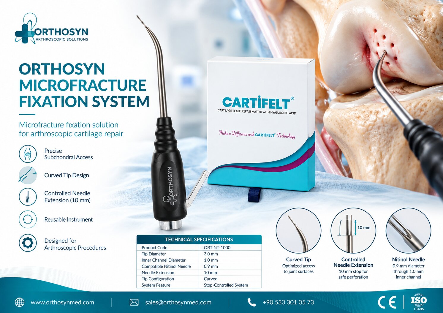
Within the ORTHOSYN arthroscopy portfolio, the ORTHOSYN Microfracture System provides instrumentation for controlled subchondral bone perforation during appropriate cartilage repair procedures.
Articular cartilage is a specialized tissue covering the ends of bones within synovial joints.
In the knee, cartilage helps provide a smooth, low-friction surface that supports joint movement and distributes mechanical loads.
Cartilage injuries may involve:
Femoral condyles
Trochlear groove
Patellar articular surface
Other weight-bearing or non-weight-bearing joint regions
A focal cartilage defect refers to a localized area of cartilage damage.
Depending on its severity, the injury may involve only the cartilage or extend into the underlying subchondral bone.
Common causes include traumatic injuries, repetitive mechanical loading, sports-related damage, and osteochondral disorders.
Because mature articular cartilage has limited intrinsic healing capacity, certain defects may require surgical intervention.
Microfracture surgery is a bone marrow stimulation technique used to treat selected full-thickness cartilage defects.
The procedure involves creating small perforations in the subchondral bone beneath the damaged cartilage.
These perforations allow blood and bone marrow elements to enter the defect.
The resulting marrow-derived clot provides an environment for the formation of repair tissue.
The simplified biological pathway is:
Cartilage Defect → Subchondral Perforation → Bone Marrow Access → Repair Clot Formation → Cartilage Repair Tissue
The repair tissue is commonly fibrocartilaginous rather than identical to the original hyaline articular cartilage.
This distinction is important because fibrocartilage and native hyaline cartilage have different structural and mechanical properties.
Microfracture is commonly performed arthroscopically.
The procedure generally involves several stages.
The surgeon examines the knee joint and identifies the cartilage defect.
Important characteristics include the defect's location, dimensions, depth, and surrounding cartilage quality.
Associated meniscal, ligamentous, or alignment abnormalities may also influence treatment planning.
Unstable cartilage fragments are removed from the defect.
The surgeon prepares the defect to establish stable cartilage margins while preserving appropriate surrounding tissue.
The calcified cartilage layer may be addressed according to the selected technique.
Small perforations are created in the subchondral bone.
The objective is to access marrow elements without causing unnecessary damage to the underlying bone.
Perforation depth, spacing, and instrument selection depend on the surgical technique and instrumentation.
Blood and marrow elements enter the defect through the perforations.
A marrow-derived clot forms over the treated area.
During healing, marrow-derived cells participate in the formation of repair tissue.
The resulting tissue may help cover the cartilage defect, although its structure and mechanical characteristics differ from those of native hyaline cartilage.
OATS (Osteochondral Autograft Transfer System) is a cartilage restoration technique that transfers osteochondral tissue from one area of the patient's joint to another.
An osteochondral graft contains two important components:
Articular cartilage
Underlying subchondral bone
During the procedure, cylindrical osteochondral plugs are harvested from a selected donor region, usually a relatively low-load-bearing area of the joint.
The plugs are then transferred into prepared recipient sockets within the cartilage defect.
The concept is:
Healthy Osteochondral Tissue → Graft Harvesting → Recipient Socket Preparation → Osteochondral Graft Transfer
Unlike microfracture, OATS introduces mature hyaline cartilage tissue directly into the treated defect.
However, the procedure also involves donor-site considerations and requires successful integration of the transferred osteochondral graft.
OATS may be performed using arthroscopic or open techniques, depending on the defect location, size, and surgical requirements.
The surgeon evaluates the cartilage defect and determines whether osteochondral autograft transplantation is appropriate.
The dimensions and depth of the defect are particularly important.
The damaged region is prepared to receive one or more cylindrical osteochondral grafts.
A socket is created according to the graft dimensions.
A cylindrical plug containing healthy cartilage and subchondral bone is harvested from a selected donor site.
The graft must be handled carefully to preserve its articular surface.
The osteochondral plug is inserted into the recipient socket.
The surgeon aims to restore an appropriate articular surface contour.
The graft is assessed for positioning and mechanical stability.
Over time, integration of the graft's bony component with the surrounding recipient bone is an important part of the healing process.
The most important difference between microfracture and OATS is the type of cartilage restoration mechanism.
Microfracture stimulates the body's repair response.
OATS transfers existing osteochondral tissue.
Characteristic | Microfracture | OATS |
|---|---|---|
Surgical principle | Bone marrow stimulation | Osteochondral autograft transplantation |
Tissue source | Patient's bone marrow | Patient's own cartilage and bone |
Cartilage outcome | Primarily fibrocartilaginous repair tissue | Transferred mature hyaline cartilage |
Bone perforation | Required | Recipient socket preparation required |
Graft harvesting | Not required | Required |
Donor-site morbidity | No osteochondral donor site | Possible |
Common surgical approach | Arthroscopic | Arthroscopic or open |
Typical defect consideration | Selected small, contained full-thickness defects | Selected focal osteochondral or chondral defects |
Main limitation | Repair tissue durability | Limited donor tissue and graft-related considerations |
Neither procedure is appropriate for every cartilage defect.
Treatment selection requires an assessment of the patient's anatomy, defect characteristics, activity demands, and overall joint condition.
The tissue formed after microfracture is generally described as fibrocartilaginous repair tissue.
Native articular cartilage is predominantly hyaline cartilage.
These tissues differ in their extracellular matrix composition and mechanical behavior.
Hyaline cartilage is specialized for joint articulation and load distribution.
Fibrocartilage contains a different collagen composition and may have different mechanical properties under repetitive joint loading.
This is one reason why long-term durability is an important consideration in microfracture procedures.
However, clinical outcomes cannot be predicted solely from the type of repair tissue.
Defect characteristics, rehabilitation, patient activity, and other factors also influence results.
OATS transfers a composite graft containing cartilage and underlying bone.
This approach provides a mature articular cartilage surface supported by a bony component.
The graft can potentially restore both cartilage and underlying bone in selected osteochondral defects.
The structure can be represented as:
Articular Cartilage Layer + Subchondral Bone = Osteochondral Graft
Successful treatment depends on appropriate graft positioning, surface congruity, stability, and biological integration.
OATS also has limitations because harvesting tissue from another joint region creates a donor site.
The available amount of suitable donor tissue is limited.
Both microfracture and OATS may be considered for selected small focal cartilage defects.
Microfracture has historically been used for relatively small, contained full-thickness cartilage lesions, particularly in appropriately selected patients.
OATS may also be considered for small or moderate focal defects, particularly when the surgeon aims to transfer an osteochondral unit containing mature hyaline cartilage.
However, there is no single defect-size threshold that determines the correct procedure for every patient.
Important considerations include:
Defect surface area
Defect depth
Subchondral bone condition
Lesion location
Patient age
Activity level
Previous cartilage surgery
Joint alignment
Meniscal and ligament stability
Defect size should therefore be evaluated together with the patient's complete clinical presentation.
Microfracture may be less predictable in larger cartilage defects, particularly in patients with high mechanical demands.
Larger defects create a greater repair area and may expose the newly formed tissue to substantial joint loading.
For this reason, other cartilage restoration procedures may be considered depending on the defect.
These can include osteochondral grafting, osteochondral allograft transplantation, or cell-based cartilage restoration techniques.
The appropriate choice depends on the specific lesion and clinical circumstances.
Potential procedural advantages include:
Arthroscopic accessibility
No requirement for osteochondral graft harvesting
Use of the patient's own marrow-derived repair response
Relatively straightforward instrumentation
Applicability to selected focal cartilage lesions
Microfracture can be performed using specialized awls, picks, or needle-based perforation instruments.
However, its limitations include the characteristics of fibrocartilaginous repair tissue and the possibility of less durable results in certain patient populations.
Potential advantages of osteochondral autograft transplantation include:
Transfer of mature hyaline articular cartilage
Restoration of the cartilage-bone unit
Applicability to selected osteochondral defects
Immediate placement of structural osteochondral tissue
Avoidance of relying exclusively on marrow-derived fibrocartilaginous repair
Important limitations include donor-site morbidity, limited graft availability, technical complexity, and the need for appropriate graft integration.
These considerations must be balanced against the expected benefits in each patient.
Microfracture does not reproduce the original hyaline cartilage structure in a predictable manner.
The repair tissue may be less resistant to repetitive mechanical loading.
Other concerns include subchondral bone changes, incomplete defect filling, and deterioration of clinical results over time in some patients.
OATS requires harvesting osteochondral tissue from another region of the joint.
This creates potential donor-site morbidity.
Additional challenges include graft positioning, surface matching, limited donor tissue availability, and integration of the graft with the surrounding bone.
Neither procedure eliminates all biological and mechanical challenges associated with cartilage restoration.
Patient activity level is an important factor when evaluating cartilage restoration.
Athletes and physically active patients may expose the repaired cartilage surface to substantial repetitive loading.
The surgeon must consider:
Sports participation
Occupational demands
Expected joint loading
Rehabilitation requirements
Defect location
Previous injuries
Associated joint instability
The decision should be individualized rather than based exclusively on whether the patient participates in sports.
Subchondral bone is the layer of bone located beneath articular cartilage.
It provides structural support for the cartilage surface and participates in the mechanical behavior of the joint.
Both microfracture and OATS involve the subchondral region, but in different ways.
In microfracture, the surgeon creates controlled perforations to access marrow elements.
In OATS, a cylindrical osteochondral graft containing subchondral bone is transferred into a prepared socket.
Therefore:
Microfracture → Subchondral Bone Perforation
OATS → Osteochondral Bone-and-Cartilage Transfer
This difference is central to understanding the biological mechanisms of the two procedures.
Microfracture requires instrumentation capable of accessing and perforating the subchondral bone.
Depending on the technique, surgeons may use:
Arthroscopic visualization equipment
Cartilage preparation instruments
Microfracture awls
Microfracture picks
Needle-based perforation systems
Depth-control instruments
Instrument selection influences surgical access, perforation control, and the ability to reach difficult anatomical regions.
Controlled perforation is particularly relevant when treating cartilage defects in confined joint spaces.
The ORTHOSYN Microfracture System is designed to provide controlled needle-based subchondral bone perforation during applicable arthroscopic cartilage repair procedures.
Its technical characteristics include:
Specification | ORTHOSYN Microfracture System |
|---|---|
Product code | ORT-NT-1000 |
Tip configuration | Curved |
Tip diameter | 3.0 mm |
Inner channel diameter | 1.0 mm |
Nitinol needle diameter | 0.9 mm |
Controlled needle extension | 10 mm |
The system incorporates a curved-tip configuration and a nitinol needle mechanism intended for controlled perforation.
The needle and stopper configuration supports defined extension during the procedure.
These features are relevant to the instrumentation requirements of microfracture techniques, where controlled access to subchondral bone is important.
The product must be used according to its applicable indications, technical documentation, and instructions for use.
During microfracture, the objective is to create perforations that provide access to marrow elements while limiting unnecessary damage to the surrounding subchondral structure.
Instrument control is important for:
Perforation positioning
Access to curved joint surfaces
Needle advancement
Reproducible technique
Management of confined anatomical spaces
The ORTHOSYN Microfracture System incorporates a controlled needle-extension concept to support the perforation procedure.
However, the clinical outcome depends on the complete surgical technique and patient-specific factors, not solely on the instrumentation.
The ORTHOSYN Microfracture System is associated with the subchondral perforation principle used in microfracture procedures.
OATS requires a different instrumentation approach involving osteochondral graft harvesting and recipient socket preparation.
These techniques should not be confused.
Microfracture instrumentation is not equivalent to an osteochondral graft transfer system.
The ORTHOSYN Microfracture System should not be presented as an OATS harvesting or transplantation device.
Rehabilitation is an important part of both procedures.
Protocols depend on the lesion location, defect size, surgical technique, and patient characteristics.
After microfracture, rehabilitation aims to protect the developing repair tissue while gradually restoring joint motion and function.
After OATS, rehabilitation must consider graft stability, bone integration, and the restoration of joint function.
Both approaches may involve:
Temporary weight-bearing restrictions
Controlled range-of-motion exercises
Progressive strengthening
Functional rehabilitation
Gradual return to sports or demanding activities
Rehabilitation protocols should be determined by the treating surgeon and rehabilitation team.
There is no universally superior procedure for every patient.
Comparative studies have reported differences in outcomes between microfracture and osteochondral autograft transplantation, particularly in certain young or highly active patient populations.
However, the interpretation of these findings depends on defect characteristics, study design, follow-up duration, and patient selection.
OATS may offer advantages in selected cases where restoration with mature osteochondral tissue is desired.
Microfracture remains an established marrow stimulation option for appropriately selected focal cartilage defects.
The correct treatment is determined by clinical evaluation rather than a universal ranking of surgical techniques.
When comparing these techniques, the surgeon may consider:
Defect size and location
Depth of cartilage damage
Subchondral bone involvement
Patient age and activity level
Quality of surrounding cartilage
Previous cartilage procedures
Joint alignment and stability
Donor-site availability
Rehabilitation requirements
Expected mechanical loading
These factors help determine whether marrow stimulation, osteochondral autograft transfer, or another cartilage restoration procedure is appropriate.
ORTHOSYN provides arthroscopic technologies intended to support selected orthopedic procedures.
Within its cartilage-related instrumentation portfolio, the ORTHOSYN Microfracture System offers a needle-based approach to controlled subchondral perforation.
The system is relevant to the following surgical concept:
Arthroscopic Cartilage Defect Assessment → Defect Preparation → Controlled Subchondral Perforation → Marrow Stimulation
ORTHOSYN focuses on providing specialized arthroscopic instrumentation and orthopedic solutions for healthcare professionals.
Further information about ORTHOSYN products is available at orthosynmed.com.
Microfracture and OATS are two different surgical approaches for treating selected focal knee cartilage defects.
Microfracture uses controlled subchondral bone perforation to stimulate a marrow-derived repair response, commonly resulting in fibrocartilaginous repair tissue.
OATS transfers cylindrical grafts containing mature hyaline cartilage and underlying bone from a donor site to the damaged region.
The fundamental distinction is:
Microfracture → Bone Marrow Stimulation → Cartilage Repair Tissue
OATS → Osteochondral Graft Transfer → Transplanted Cartilage and Bone
Both techniques have specific indications, advantages, and limitations.
Within the ORTHOSYN arthroscopy portfolio, the ORTHOSYN Microfracture System (ORT-NT-1000) provides specialized instrumentation for controlled subchondral bone perforation during applicable cartilage repair procedures.
The choice between microfracture, OATS, and other cartilage restoration techniques should always be based on individual patient assessment, defect characteristics, and the treating surgeon's clinical judgment.