


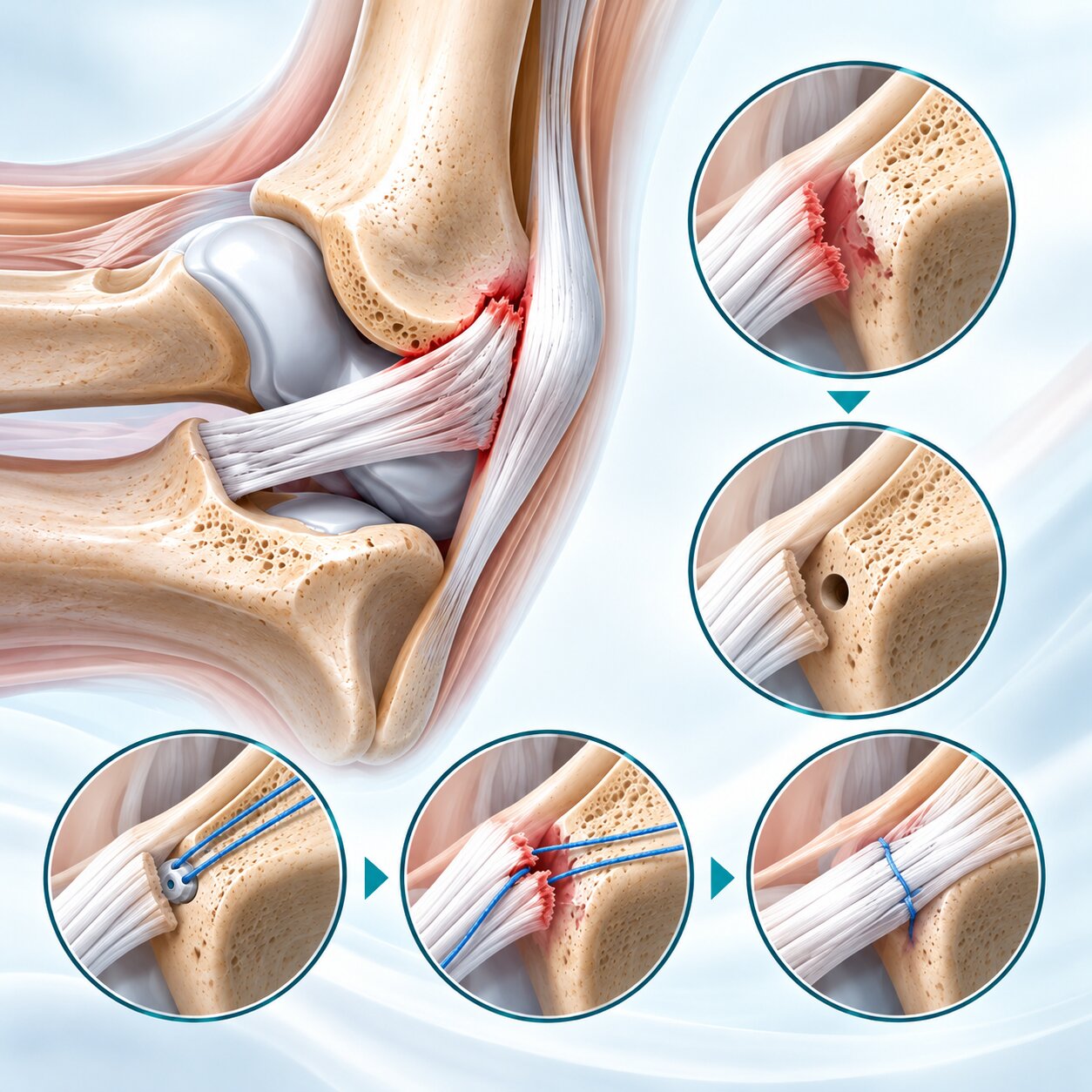
Ulnar collateral ligament (UCL) repair is a surgical technique used to restore ligament stability in selected injuries involving the medial side of the elbow. Suture anchors can play an important role in this procedure by securing damaged ligament tissue back to its anatomical attachment site on the bone.
The UCL, also known as the medial collateral ligament of the elbow, helps resist valgus stress and contributes to elbow stability during throwing and other demanding upper-limb activities.
When the ligament is detached from its humeral or ulnar attachment, suture anchors may be used to reattach the tissue to bone. Depending on the injury pattern, repair can also be combined with a suture-based internal brace augmentation technique.
Within the ORTHOSYN arthroscopy portfolio, Soft Anchors and Titanium Suture Anchors represent different soft tissue-to-bone fixation technologies. Their potential use in UCL repair must be evaluated according to the specific product indications and instructions for use.
The ulnar collateral ligament (UCL) is located on the medial, or inner, side of the elbow.
It connects structures of the distal humerus and proximal ulna and contributes to elbow stability.
The UCL complex is commonly described as having three components:
Anterior bundle
Posterior bundle
Transverse bundle
The anterior bundle is particularly important for resisting valgus stress during elbow movement.
This function is especially relevant in overhead throwing sports, where substantial forces act across the medial elbow.
UCL injuries can occur through repetitive mechanical loading or acute trauma.
Common contributing factors include:
Repetitive overhead throwing
Baseball pitching
Javelin throwing
High valgus loading
Sudden traumatic elbow injuries
Contact sports
Repetitive stress during athletic activity
In throwing athletes, repeated valgus loading can contribute to progressive ligament damage.
Acute UCL injuries may also occur when a sudden force exceeds the ligament's capacity to resist loading.
Symptoms depend on the location, severity and chronicity of the injury.
Patients may experience:
Pain along the inner elbow
Discomfort during throwing
Reduced throwing performance
A sensation of elbow instability
Tenderness near the medial elbow
Pain during valgus stress
Reduced confidence during athletic activity
Some patients report a sudden popping sensation during an acute injury.
Clinical examination and imaging are used to determine the injury pattern and appropriate treatment.
Diagnosis generally involves a combination of patient history, physical examination and imaging.
Clinical assessment may include:
Medial elbow tenderness
Valgus stress testing
Moving valgus stress test
Range-of-motion assessment
Evaluation of the ulnar nerve
Assessment of associated elbow pathology
MRI, sometimes with arthrographic techniques, can help evaluate ligament integrity, tear location and tissue condition.
These findings are important when deciding whether a ligament is suitable for repair or requires reconstruction.
UCL repair and reconstruction are different surgical approaches.
UCL repair aims to preserve and reattach the patient's existing ligament tissue.
UCL reconstruction, commonly associated with Tommy John surgery, uses a tendon graft to replace the function of an insufficient ligament.
Characteristic | UCL Repair | UCL Reconstruction |
|---|---|---|
Main objective | Reattach existing ligament | Reconstruct ligament using a graft |
Tissue requirement | Adequate native ligament quality | Graft tissue |
Common fixation concept | Suture anchors | Graft fixation through tunnels or other constructs |
Typical consideration | Selected acute or repairable avulsion injuries | Poor tissue quality or injuries unsuitable for repair |
Internal brace augmentation | May be used | Not inherent to conventional reconstruction |
The decision depends on tear location, ligament quality, injury history, patient requirements and surgeon assessment.
UCL repair may be considered in selected patients with injuries that have suitable tissue quality and a repairable configuration.
Examples include certain proximal or distal ligament avulsions.
Important factors include:
Tear location
Ligament tissue quality
Acute versus chronic injury
Degree of ligament degeneration
Associated elbow injuries
Patient activity level
Athletic demands
Not every UCL tear is appropriate for direct repair.
Chronic midsubstance degeneration or extensive ligament damage may require a different surgical strategy.
A suture anchor is an orthopedic fixation device designed to secure sutures to bone.
In soft tissue repair, the anchor provides a fixation point while the sutures capture and stabilize the tissue.
The basic concept is:
Bone → Suture Anchor → High-Strength Suture → Ligament
Suture anchors are used in various orthopedic procedures involving soft tissue-to-bone fixation.
Depending on the product design, anchors may be manufactured from titanium, PEEK or high-strength textile materials.
In selected UCL injuries, a ligament may detach from its anatomical attachment site.
The surgical objective is to restore the ligament's connection to bone.
A suture anchor is positioned at an appropriate fixation site, and the attached sutures are passed through or around the ligament tissue.
The sutures are then secured to maintain the ligament near its intended attachment.
A simplified fixation pathway is:
Humerus or Ulna → Suture Anchor → Suture → UCL Tissue
The procedure may involve one or more fixation points depending on the tear pattern and surgical technique.
The surgeon evaluates the damaged ligament and surrounding structures.
Important considerations include:
Ligament attachment site
Tear configuration
Tissue quality
Associated injury
Ulnar nerve anatomy
Bone quality
The surgeon determines whether the ligament can be adequately repaired.
When the ligament has detached from bone, the relevant attachment site is identified.
The bone surface may be prepared according to the surgical technique to create an appropriate environment for reattachment.
The objective is to restore the relationship between the ligament and its anatomical attachment.
A suture anchor is inserted into the selected bone fixation site.
Depending on the location of the tear, fixation may involve the humeral or ulnar attachment region.
The anchor provides a stable point for the suture construct.
The concept is:
Bone Fixation Site → Anchor Placement → Suture Availability
Anchor size, drilling requirements and insertion technique depend on the specific implant system and local anatomy.
The sutures connected to the anchor are passed through the damaged ligament tissue.
Suture configuration depends on the technique and tissue quality.
The objective is to capture sufficient ligament tissue while avoiding unnecessary tissue damage.
A simplified representation is:
Anchor → Suture Passage → Ligament Capture
The sutures are tensioned and secured to position the ligament against its intended attachment site.
The surgeon evaluates the repair construct and its relationship to the surrounding anatomy.
The fixation concept becomes:
Bone → Anchor → Suture → Reattached UCL
The objective is to maintain the ligament in an appropriate position during the healing process.
After fixation, the surgeon evaluates elbow stability and the repair construct.
This assessment may include:
Ligament tension
Elbow range of motion
Valgus stability
Suture security
Fixation integrity
The final repair should follow the selected surgical technique and implant-specific instructions.
Internal brace augmentation is a technique in which a high-strength suture tape or similar reinforcement construct is used alongside a repaired ligament.
The concept is to provide additional mechanical support to the repair during the healing period.
A simplified configuration is:
Bone Anchor → Suture Tape Reinforcement → Bone Anchor
The native ligament is repaired while the augmentation construct spans the relevant attachment sites.
Internal brace augmentation is not the same as ligament reconstruction with a tendon graft.
Its suitability depends on the injury pattern, fixation system and surgical technique.
Importantly, not every suture anchor is approved or designed for internal brace augmentation. Product-specific compatibility and indications must be confirmed.
The medial elbow contains relatively compact anatomical structures.
Anchor dimensions may therefore be important when planning fixation.
Small-diameter anchors can be relevant to procedures requiring fixation within limited bone dimensions.
Potential considerations include:
Bone tunnel diameter
Available bone stock
Anchor positioning
Ligament attachment anatomy
Multiple fixation points
Proximity to important anatomical structures
However, smaller diameter alone does not establish that an anchor is appropriate for a particular UCL repair technique.
The anchor must provide suitable fixation performance and be used according to its intended purpose.
Soft Anchors, also called all-suture anchors, use high-strength textile-based fixation constructs rather than conventional rigid anchor bodies.
These devices are designed around a soft tissue-to-bone fixation principle.
Potential characteristics of soft anchor technology include:
Small insertion diameter
Textile-based anchor construction
Reduced rigid implant material
Compatibility with selected minimally invasive techniques
High-strength suture fixation concepts
The mechanical performance of an all-suture anchor depends on its design, deployment, bone quality and loading conditions.
Within the ORTHOSYN arthroscopy portfolio, ORTHOSYN Soft Anchors are manufactured using UHMWPE-based textile technology.
Available diameters include:
Anchor diameter | ORTHOSYN Soft Anchor |
|---|---|
1.3 mm | Soft Anchor |
1.5 mm | Soft Anchor |
1.9 mm | Soft Anchor |
3.0 mm | Soft Anchor |
Configurations with and without needles are available.
These devices provide small-diameter soft tissue fixation technologies within the ORTHOSYN anchor portfolio.
For UCL repair, the suitability of any specific Soft Anchor model must be confirmed against its documented indications, fixation requirements and instructions for use.
Titanium suture anchors provide a rigid metallic fixation option.
A typical titanium anchor uses a threaded or otherwise mechanically engaging design to establish fixation within bone.
Sutures attached to the anchor are used to secure the ligament or other soft tissue.
The fixation concept is:
Titanium Anchor → Bone Fixation → High-Strength Suture → Ligament Reattachment
Titanium anchors are relevant to soft tissue-to-bone fixation procedures where the implant's dimensions and intended use are appropriate.
ORTHOSYN offers titanium suture anchor technologies in several diameters, including:
2.0 mm
3.5 mm
5.0 mm
6.5 mm
The appropriate anchor size depends on the anatomical region, bone characteristics and surgical technique.
For UCL repair, particular attention must be given to the available bone dimensions and the product's documented intended purpose.
The existence of a particular anchor size does not establish its suitability for every ligament repair application.
Soft and titanium anchors use different structural concepts.
Characteristic | Soft Anchor | Titanium Anchor |
|---|---|---|
Anchor body | Textile-based | Metallic |
Fixation concept | Soft anchor deployment within bone | Rigid anchor engagement with bone |
Material | UHMWPE-based textile | Titanium |
Implant structure | Flexible | Rigid |
Available ORTHOSYN diameters | 1.3–3.0 mm | 2.0–6.5 mm |
UCL repair suitability | Requires product-specific confirmation | Requires product-specific confirmation |
Neither anchor category should be considered universally superior.
Fixation selection depends on anatomy, bone quality, tissue characteristics, surgical technique and implant-specific evidence.
The suture forms the mechanical connection between the anchor and ligament tissue.
Important considerations include:
Tensile strength
Suture configuration
Tissue capture
Knot security, where applicable
Resistance to abrasion
Tensioning
Fixation stability
UHMWPE-based sutures are commonly used in orthopedic soft tissue fixation because of their high-strength characteristics.
Within the ORTHOSYN portfolio, Enhanced Sutures provide UHMWPE-based braided suture technology for relevant orthopedic fixation applications.
Their use in a specific UCL repair construct must follow applicable product indications and compatibility requirements.
The UCL contributes to the stability of the medial elbow.
When repair is selected, the objective is to restore the damaged ligament as closely as appropriate to its anatomical attachment.
Proper positioning is important because the ligament's function depends on its relationship to the humerus, ulna and elbow joint.
The repair concept is:
Anatomical Attachment → Ligament Reattachment → Mechanical Stabilization → Biological Healing
However, anatomical repair alone does not guarantee successful healing or return to sport.
Outcomes also depend on tissue quality, fixation integrity, rehabilitation and patient-related factors.
UCL injuries are particularly relevant in overhead throwing sports.
Baseball pitchers are frequently discussed because the throwing motion produces substantial valgus forces across the medial elbow.
Other relevant sports include:
Baseball
Softball
Javelin
Handball
Tennis
Other overhead throwing activities
When a UCL injury requires surgery, the decision between repair and reconstruction depends on the specific ligament injury.
The educational pathway can be represented as:
Throwing-Related Elbow Pain → UCL Injury → Clinical Evaluation → Repair or Reconstruction → Ligament Fixation
Within the ORTHOSYN arthroscopy portfolio, several product technologies are relevant to the broader principles of soft tissue-to-bone fixation.
These include:
ORTHOSYN Soft Anchors
UHMWPE-based textile anchors available in small diameters.
ORTHOSYN Titanium Suture Anchors
Rigid metallic anchors designed for selected soft tissue fixation applications.
ORTHOSYN Enhanced Sutures
High-strength UHMWPE-based braided sutures for applicable orthopedic procedures.
Together, these technologies represent different approaches to:
Bone Fixation → Suture Connection → Soft Tissue Stabilization
Their potential use in UCL repair must be confirmed through the specific product's approved indications, instructions for use and supporting technical documentation.
Explore the ORTHOSYN Suture Anchors portfolio for further product information.
Suture anchors are used in selected UCL repair procedures to secure damaged ligament tissue back to its anatomical bone attachment.
The anchor establishes a fixation point within the humerus or ulna, while the attached sutures capture and stabilize the ligament.
The fundamental concept is:
Bone → Suture Anchor → High-Strength Suture → UCL Reattachment
In selected cases, ligament repair may also be combined with internal brace augmentation.
Within the ORTHOSYN arthroscopy portfolio, Soft Anchors, Titanium Suture Anchors and Enhanced Sutures provide technologies relevant to the broader principles of soft tissue-to-bone fixation.
The suitability of individual ORTHOSYN products for UCL repair must be established through their documented intended purposes and instructions for use.