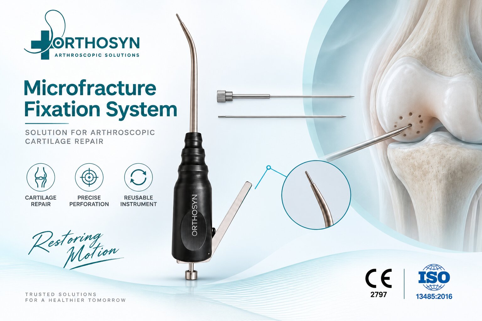
Articular cartilage defects of the knee can develop after traumatic injuries, repetitive mechanical stress, or focal damage to the joint surface. Because articular cartilage has a limited intrinsic healing capacity, selected focal cartilage lesions may require surgical treatment.
One arthroscopic technique used for this purpose is microfracture surgery. The procedure involves creating small perforations in the subchondral bone beneath the cartilage defect to access the underlying bone marrow and support the formation of repair tissue.
Microfracture therefore represents an important area of knee arthroscopy, cartilage repair and sports medicine.
Articular cartilage covers the surfaces of the bones within the knee joint and provides a smooth, low-friction surface for movement.
A focal cartilage defect occurs when a localized area of this cartilage is damaged.
Cartilage defects may involve different areas of the knee, including:
The size, depth and location of the defect, together with patient-related factors, influence the treatment strategy.
Microfracture surgery is a bone marrow stimulation technique used for selected articular cartilage defects.
After the damaged area has been identified arthroscopically, unstable or damaged cartilage is prepared to create a defined defect.
Small perforations are then created in the exposed subchondral bone.
These perforations allow blood and bone marrow components to reach the treated cartilage defect.
The basic concept can be summarized as:
Cartilage Defect → Defect Preparation → Subchondral Bone Perforation → Bone Marrow Access → Repair Response
Microfracture can be performed during knee arthroscopy, allowing the surgeon to visualize the cartilage lesion and access the affected joint surface.
The general procedure involves:
The exact number, depth and distribution of perforations depend on the lesion and surgical technique.
Articular cartilage itself has limited vascularity and therefore limited intrinsic healing potential.
Microfracture attempts to stimulate a biological repair response by creating controlled access to the subchondral bone marrow.
Blood and marrow components can enter the prepared defect through the microperforations.
This is why microfracture is classified as a bone marrow stimulation technique rather than simply a mechanical cartilage preparation procedure.
The procedure requires instruments capable of reaching the cartilage defect and creating controlled perforations in the subchondral bone.
Depending on the anatomical location of the lesion, instrument geometry can be important for obtaining the appropriate approach angle.
Dedicated microfracture awls or cannulas can therefore be used during arthroscopic cartilage procedures.
ORTHOSYN provides a Microfracture Cannula designed for arthroscopic applications and controlled access to the treatment area.
Cartilage defects can occur on curved and anatomically difficult-to-access joint surfaces.
The instrument must therefore approach the subchondral surface at an appropriate orientation to create the intended perforation.
This is particularly relevant when treating lesions involving different regions of the:
Instrument design and surgical access therefore play an important role in arthroscopic microfracture procedures.
Microfracture is one of several surgical strategies used for cartilage defects.
Other cartilage restoration approaches may include:
These techniques do not have identical indications.
The appropriate treatment depends on factors such as:
Microfracture should therefore be considered within the broader field of cartilage repair and restoration, rather than as a universal treatment for every cartilage lesion.
Yes. Focal cartilage defects can occur in physically active patients and athletes following traumatic or repetitive joint injuries.
For this reason, searches related to cartilage damage frequently overlap with sports medicine and knee arthroscopy.
The search pathway can be considered as:
Sports Injury → Knee Cartilage Damage → Focal Cartilage Defect → Arthroscopic Cartilage Repair → Microfracture Surgery
This gives the topic broader SEO value than focusing only on the surgical instrument itself.
ORTHOSYN's arthroscopy portfolio includes instruments used in procedures beyond ligament reconstruction and meniscal repair.
The ORTHOSYN Microfracture Cannula provides an instrument solution for arthroscopic procedures involving controlled access to the subchondral bone.
This expands the ORTHOSYN arthroscopy portfolio into another important area of knee surgery:
ACL/PCL Reconstruction → Meniscal Repair → Cartilage Repair → Microfracture
As a result, the portfolio addresses multiple stages and indications within arthroscopic sports medicine rather than focusing exclusively on ligament fixation.
Microfracture surgery is an arthroscopic bone marrow stimulation technique used for selected focal articular cartilage defects.
The procedure involves preparing the cartilage lesion and creating controlled microperforations in the subchondral bone to provide access to underlying blood and bone marrow components.
Because successful execution requires appropriate access to the defect and controlled perforation of the subchondral surface, dedicated microfracture instruments play an important role in the procedure.
For ORTHOSYN, this topic also expands our SEO coverage beyond anchors, ACL reconstruction and meniscal repair into knee cartilage repair, cartilage defects, bone marrow stimulation and sports medicine.